Oral Abstracts Session
Virtual Recording

Andrew D. Scott, PhD, FSCMR
Associate Professor
Imperial College London and Royal Brompton Hospital
London, England, United Kingdom
Andrew D. Scott, PhD, FSCMR
Associate Professor
Imperial College London and Royal Brompton Hospital
London, England, United Kingdom
Yaqing Luo
PhD Student
Royal Brompton Hospital and National Heart and Lung Institute, Imperial College London
London, England, United Kingdom
.jpg "Pedro F. Ferreira, PhD photo")
Pedro F. Ferreira, PhD
CMR Physicist
Royal Brompton Hospital
London, England, United Kingdom
Alberto Di Biase, MSc
Research Assistant
Imperial College London
Lonodn, England, United Kingdom
.jpg "Karl P. Kunze, PhD photo")
Karl P. Kunze, PhD
Senior Cardiac MR Scientist
Siemens Healthineers
Camberley, England, United Kingdom

Dudley Pennell, MD, FSCMR
Director of cardiac MRI
Royal Brompton Hospital
London, England, United Kingdom

Sonia Nielles-Vallespin, PhD, MSc, BSc
Senior Lecturer
Imperial College London
London, England, United States
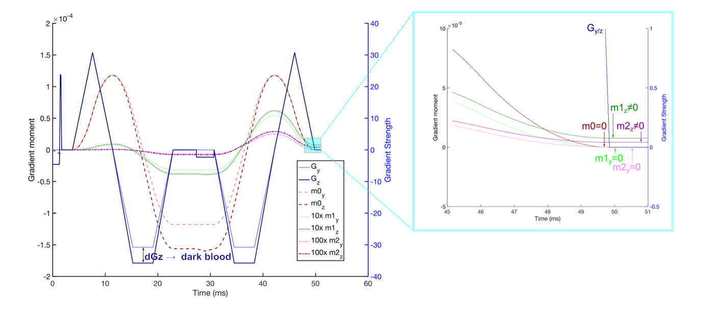 Figure 2: Optimising the gradient offset used to minimise blood signal in the b=50smm-2 diffusion weighted images (higher b-value images typically have less residual blood signal). The original second order motion compensated images include substantial residual blood signal (top right), making segmentation of the endocardium and right ventricular septal border difficult without partial volume effects. Applying the gradient offset in-plane (phase direction, bottom left) results in motion induced signal loss (magenta arrows) while providing in complete blood suppression (mauve arrow). Applying the offset gradient through plane (slice direction, bottom right) with a large magnitude results in motion related signal loss.
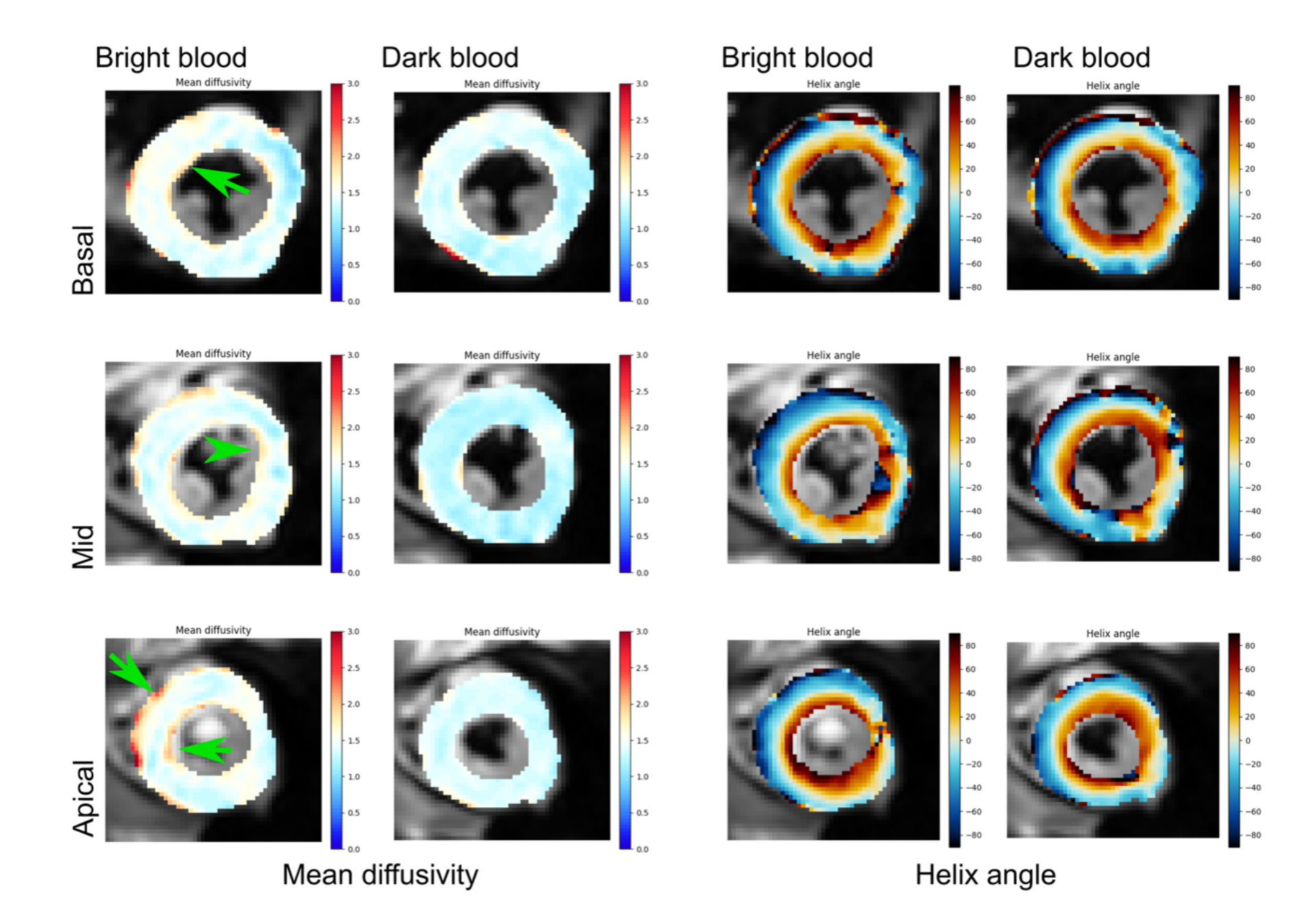
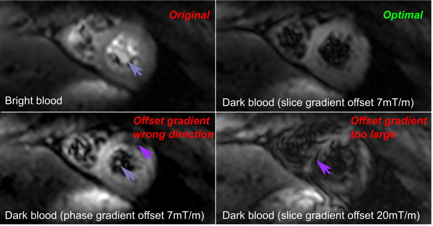 Figure 3: A comparison of mean diffusivity (MD, units x10-3mm2s-1) and helix angle (HA, units degrees) acquired in an a different example subject in three short axis slices. Even in the basal slice where the blood signal is minimal even without the dark blood offset gradient, the MD maps show increased MD values in myocardium bordering on blood pool (arrows highlight increased MD due to blood signal), which is removed with the dark blood preparation. HA maps appear similar in both cases, although the blood suppression allows endocardial regions to be analysed without blood signal contamination.