Rapid Fire Session

Christine Mancini, RT, FSCMR
CV MRI Technologist
National Heart, Lung, and Blood Institute, National Institutes of Health
Bethesda, Maryland, United States
Sant Kumar
Fellow
National Heart, Lung, and Blood Institute, National Institutes of Health, United States
W. Patricia Bandettini, MD
Senior Research Physician
National Heart, Lung, and Blood Institute, National Institutes of Health
Bethesda, Maryland, United States
Jennifer Henry
The National Institutes of Health, United States
Margaret Lowery, RN
National Heart, Lung and Blood Institute (NHLBI), United States
Scott Baute
Physician Assistant
National Heart, Lung, and Blood Institute, National Institutes of Health, United States
Anastasia Tsakirellis
Nurse Practitioner
National Institutes of Health, United States

Adrienne E E. Campbell-Washburn, PhD
Principal Investigator
National Heart, Lung, and Blood Institute, National Institutes of Health
Bethesda, Maryland, United States
| 0.55T T1 values (ms) | |||||
| Native | Post-contrast | ECV | |||
| Myocardium | Blood | Myocardium | Blood | ||
| Healthy volunteers | 704 ± 31 (61) | 1141 ± 79 (61) | 395 ± 82 (6) | 304 ± 101 (6) | 26 ± 4 (5) |
| MI (remote) | 705 ± 37 (34) | 1154 ± 63 (34) | 354 ± 38 (32) | 264 ± 30 (32) | 27 ± 5 (32) |
| MI (infarcted) | 559 ± 141 (25) | -- | 207 ± 69 (24) | -- | 64 ± 21 (24) |
| Cardiomyopathy | 717 ± 36 (19) | 1126 ± 102 (19) | 380 ± 71 (14) | 302 ± 154 (14) | 26 ± 5 (14) |
| Acute inflammatory process | 756 ± 69 (10) * | 1157 ± 113 (10) | 382 ± 47 (5) | 302 ± 82 (5) | 28 ± 6 (5) |
| Amyloid | 774 ± 26 (4) ** | 1147 ± 46 (4) | 253 ± 60 (3) | 265 ± 15 (3) | 54 ± 18 (3) |
| Hepatic iron overload | 731 ± 41 (43) | 1175 ± 83 (43) | -- | -- | -- |
| Acute COVID-19 | 719 ± 18 (16) | 1154 ± 82 (16) | 348 ± 16 (15) | 243 ± 25 (15) | 27 ± 3 (15) |
| Post-COVID-19 | 714 ± 22 (199) | 1123 ± 105 (197) | 349 ± 31 (185) | 240 ± 34 (184) | 26 ± 4 (184) |
| Other cardiovascular condition | 705 ± 28 (46) | 1145 ± 75 (46) | 362 ± 43 (30) | 256± 48 (30) | 24 ± 3 (30) |
| 1.5T T1 values (ms) | |||||
| Native | Post-contrast | ECV | |||
| Myocardium | Blood | Myocardium | Blood | ||
| Healthy volunteers | 1028 ± 34 (14) | 1592 ± 103 (14) | 544 ± 19 (3) | 413 ± 39 (3) | 27 ± 3 (3) |
| MI (remote) | 1027 ± 60 (33) | 1598 ± 31 (33) | 448 ± 68 (33) | 319 ± 30 (33) | 28 ± 6 (33) |
| MI (infarcted) | 948 ± 28 (24) | -- | 272 ± 78 (24) | -- | 61 ± 18 (24) |
| Cardiomyopathy | 1029 ± 51 (19) | 1520 ± 136 (19) | 439 ± 39 (15) | 306 ± 38 (14) | 26 ± 4 (14) |
| Acute inflammatory process | 1093 ± 74 (9) * | 1676 ± 132 (9) | 441 ± 56 (9) | 320 ± 80 (9) | 32 ± 7 (9) |
| Amyloid | 1139 ± 50 (3) * | 1607 ± 43 (3) | 296 ± 68 (3) | 303 ± 21 (3) | 54 ± 18 (3) |
| Hepatic iron overload | 1012 ± 57 (43) | 1572 ± 100 (43) | -- | -- | -- |
| Acute COVID-19 | -- | -- | -- | -- | -- |
| Post-COVID-19 | 998 ± 23 (5) | 1498 ± 79 (5) | 450 ± 18 (5) | 287 ± 17 (5) | 24 ± 2 (5) |
| Other cardiovascular conditions | 1027 ± 32 (40) | 1608 ± 87 (39) | 453 ± 34 (26) | 298 ± 39 (26) | 25 ± 2 (26) |
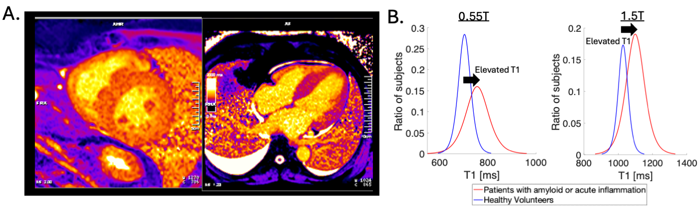
 Figure 2: A) T1 maps from a patient presenting with severe left ventricular hypertrophy and exertional dyspnea. At 0.55T, myocardial native T1 values were increased at 760-790 ms (vs normal 704ms) and the extracellular volume fraction was 64%. The patient was confirmed to have cardiac amyloidosis. B) Histograms (with logistic fit) of T1 values to demonstrate elevated T1 in patients with amyloid or acute inflammation.