
Case Session
Virtual Recording

Ankush Ankush, MD
Cardiothoracic Imaging Fellow
Atrium Wake Forest Baptist Medical Center
Winston Salem, North Carolina, United States
Muthu Sakthivel, MD, FSCMR
Assistant Professor/Cardiothoracic Imaging Fellowship Program Director
UNC School of Medicine
Chapel Hill, North Carolina, United States
Eswara Sellamuthu, MD
Assistant Professor
Atrium Health Wake Forest Baptist Medical Center, United States
Usha Jayagurunathan, MD
Assistant Professor
Atrium Health Wake Forest Baptist Medical Center, United States
HARISH GUDI, MD
Assistant Professor
University of Arizona College of Medicine, United States
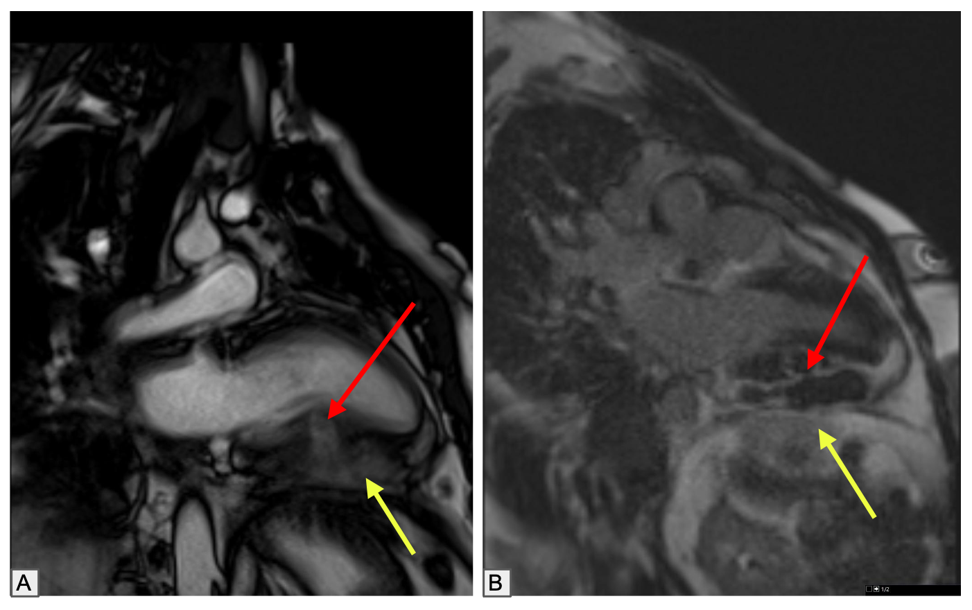 Figure 2. Cardiac MRI—LGE delineation of pericardial abscesses with myocardial involvement. (A) Short-axis phase-sensitive inversion recovery (SA PSIR) late gadolinium enhancement (LGE) images show multifocal pericardial collections with avid rim enhancement and centrally nonenhancing fluid, consistent with pericardial abscesses (white arrows). (B) SA PSIR LGE images again demonstrate pericardial rim enhancement with central nonenhancing fluid (blue arrows) and a similar focus within the inferior mid–left ventricular myocardium, compatible with a myocardial abscess (red arrow). (C) Axial post-contrast T1-weighted fat-suppressed images demonstrate multifocal, multiloculated pericardial collections with avid peripheral rim enhancement (green arrows) surrounding centrally nonenhancing fluid components (red arrows), in keeping with pericardial abscesses.
.png) Figure 3. Cardiac MRI—post-treatment response of pericardial abscess with myocardial extension. (A) Two-chamber localizer cine images obtained 4 weeks after initiation of antifungal therapy (amphotericin) show interval decrease with only trace residual pericardial collection (yellow arrow) and marked reduction of the prior myocardial extension. (B) Two-chamber PSIR late gadolinium enhancement (LGE) images at 4 weeks demonstrate decreased avid rim enhancement of the pericardial abscess (yellow arrow) with only a minimal residual myocardial component (red arrow). (C) Short-axis magnitude-reconstructed LGE images at 4 weeks show decreased extent of pericardial enhancement and reduction in the size of the multifocal, multiloculated, centrally nonenhancing fluid components.
Figure 3. Cardiac MRI—post-treatment response of pericardial abscess with myocardial extension. (A) Two-chamber localizer cine images obtained 4 weeks after initiation of antifungal therapy (amphotericin) show interval decrease with only trace residual pericardial collection (yellow arrow) and marked reduction of the prior myocardial extension. (B) Two-chamber PSIR late gadolinium enhancement (LGE) images at 4 weeks demonstrate decreased avid rim enhancement of the pericardial abscess (yellow arrow) with only a minimal residual myocardial component (red arrow). (C) Short-axis magnitude-reconstructed LGE images at 4 weeks show decreased extent of pericardial enhancement and reduction in the size of the multifocal, multiloculated, centrally nonenhancing fluid components..png)
