Quick Fire Session

Aamuktha R. Karla, MD
Internal Medicine PGY-2
Vanderbilt University Medical Center
Nashville, Tennessee, United States
Aamuktha R. Karla, MD
Internal Medicine PGY-2
Vanderbilt University Medical Center
Nashville, Tennessee, United States

Gina E. Lazzara, MD
Advanced Imaging Fellow
Vanderbilt University Medical Center
Nashville, Tennessee, United States
Satish Mishra, MD
Advanced Imaging Fellow
Vanderbilt University Medical Center, United States
Luke Laws, MD
Clinical Fellow
Vanderbilt University Medical Center, United States
John A. Wells, IV, MD
Assistant Professor of Clinical Medicine
Vanderbilt University Medical Center
Nashville, Tennessee, United States

Tania Ruiz M., MD
Assistant Professor of Medicine
Vanderbilt University Medical Center
Nashville, Tennessee, United States

Bruno B. Lima, MD, PhD
Assistant Professor of Medicine
Vanderbilt University Medical Center
Nashville, Tennessee, United States
Jeffrey M. Dendy, MD
Assistant Professor of Medicine
Vanderbilt University Medical Center
Brentwood, Tennessee, United States
Sean G. Hughes, MD, FSCMR
Associate Professor of Clinical Medicine and Radiology
Director, Cardiac MRI and Advanced Cardiac Imaging Fellowship
Vanderbilt University Medical Center
Brentwood, Tennessee, United States
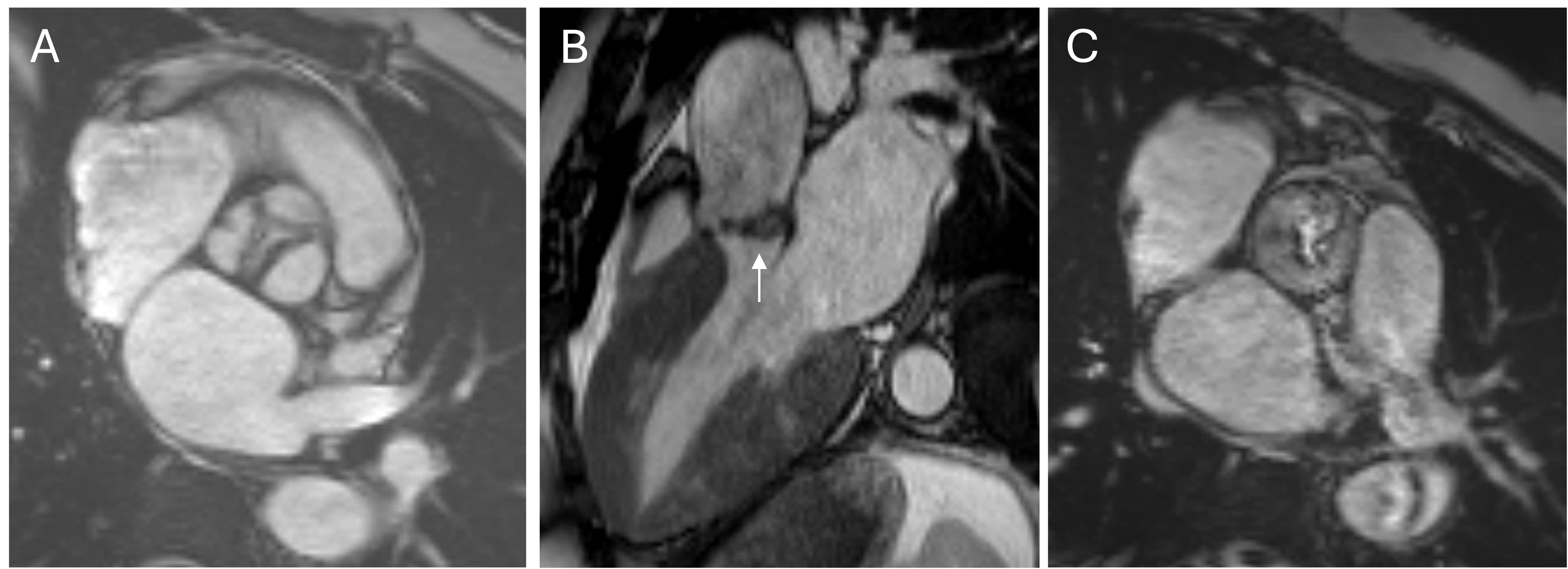 Short axis (A-C) and two-chamber (D) views demonstrating small focal subendocardial infarctions of the basal lateral and inferior walls.
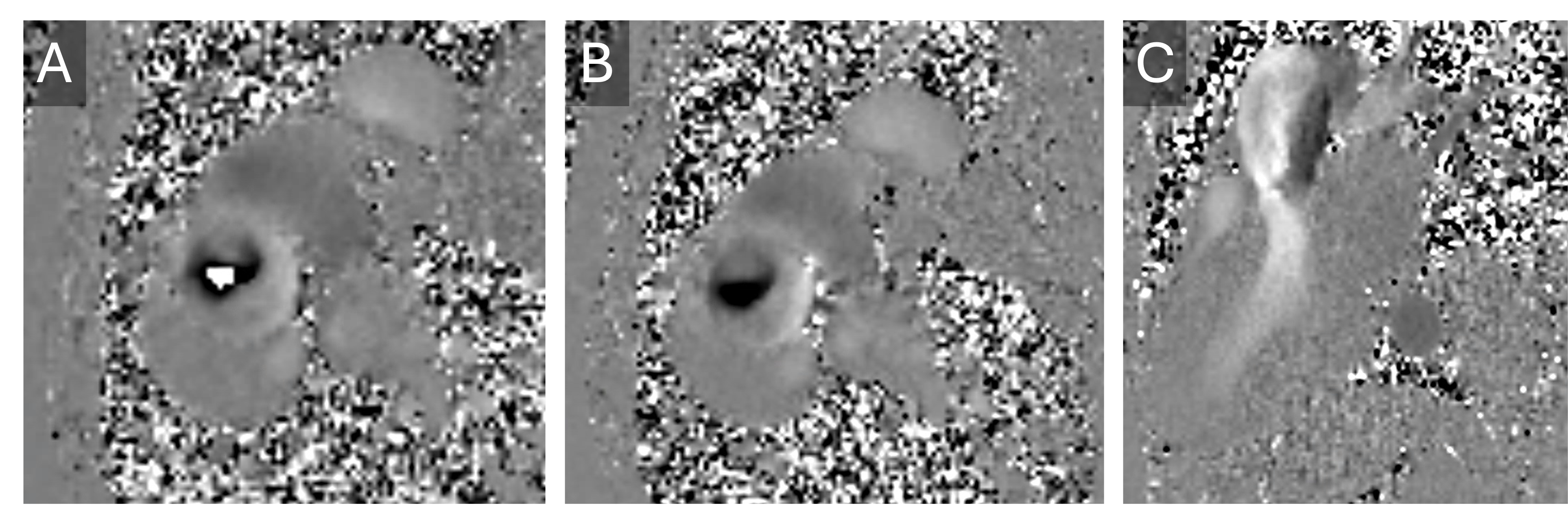
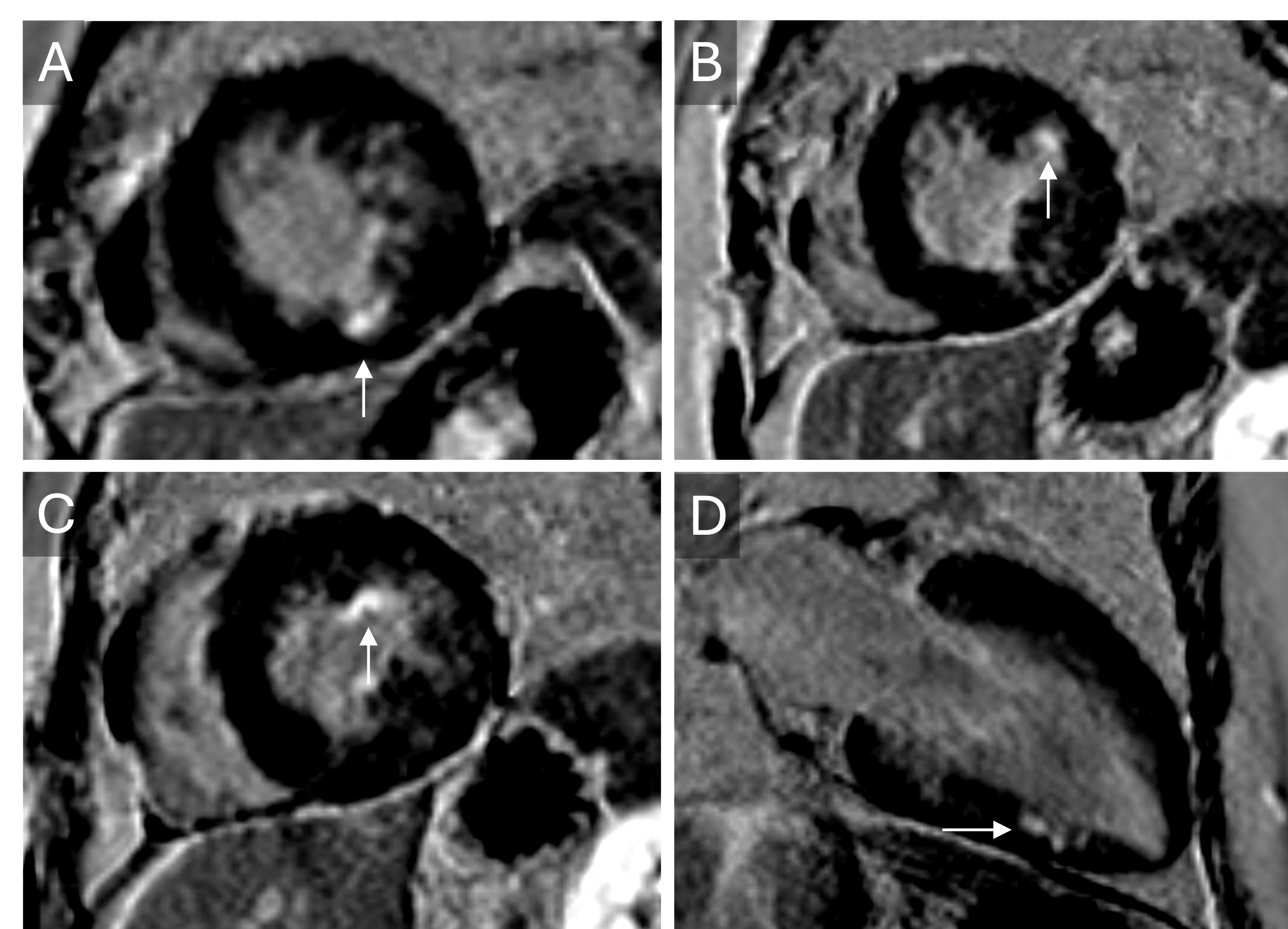 Thru-plane velocity flow mapping of the aortic valve showing aliasing at an encoding velocity of 3.25 m/s (A) that resolves at 4 m/s (B). In-plane velocity flow mapping in LVOT view (C) revealing aortic turbulence. The peak aortic velocity is estimated between 3.25 and 4.0 m/s, consistent with moderate stenosis.