Rapid Fire Session

Luuk H.G.A Hopman, PhD
Postdoctoral Researcher Cardiology
Amsterdam UMC
Amsterdam, Noord-Holland, Netherlands
Luuk H.G.A Hopman, PhD
Postdoctoral Researcher Cardiology
Amsterdam UMC
Amsterdam, Noord-Holland, Netherlands
Michiel J.B. Kemme, MD, PhD
Cardiologist-Electrophysiologist
Amsterdam Medical Center, Netherlands
Pranav Bhagirath, MD, PhD
Cardiologist
Amsterdam UMC
Amsterdam, Noord-Holland, Netherlands

Raschel Luijk
xx
Amsterdam Medical Center, Netherlands

Cor P. Allaart, MD, PhD
Cardiologist
Amsterdam Medical Center
Amsterdam, Noord-Holland, Netherlands
Marco J.W Götte, MD, PhD
MD, PhD
Stephenson Cardiac Imaging Centre
Calgary, Alberta, Canada
.jpg) Right ventricular two-chamber view (comparable fluoroscopy orientation: right anterior oblique), basal short axis view (comparable fluoroscopy orientation: left anterior oblique), and four-chamber view. The yellow arrow indicates the foramen ovale.
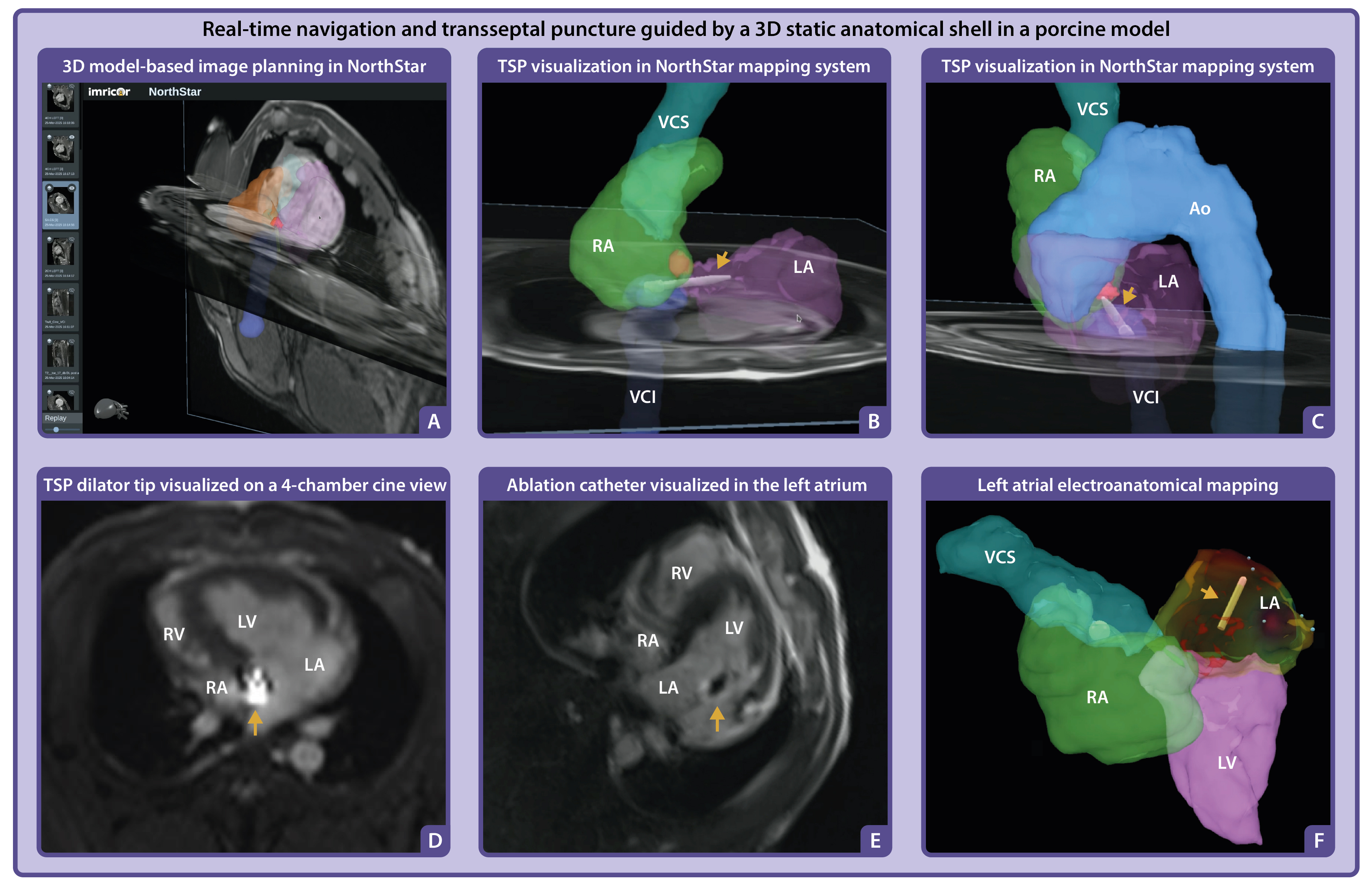
Right ventricular two-chamber view (comparable fluoroscopy orientation: right anterior oblique), basal short axis view (comparable fluoroscopy orientation: left anterior oblique), and four-chamber view. The yellow arrow indicates the foramen ovale.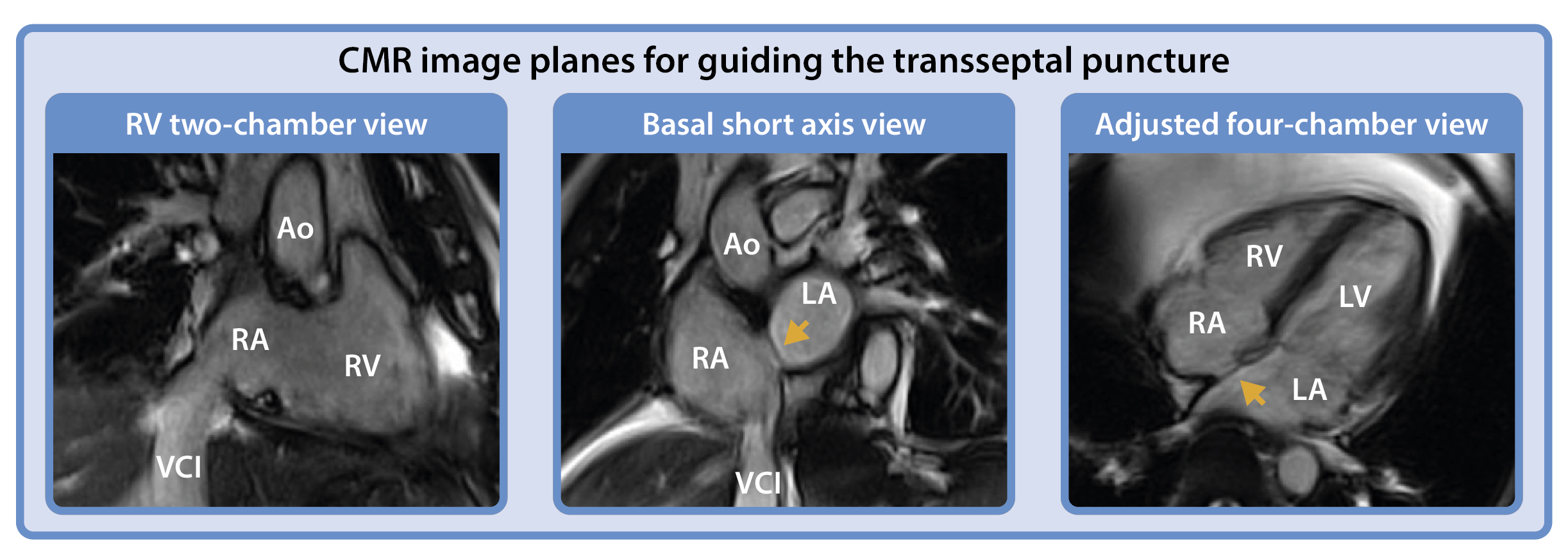 A. Navigation of the MR-compatible TSP set within a 3D static anatomical shell imported into the NorthStar Mapping System, supplemented with adjustable 2D image planes to provide additional spatial orientation and visualization of cardiac motion. B–C. The TSP set was navigated to the foramen ovale (highlighted in red segmentation), with positioning verified across multiple image planes to ensure accurate alignment for septal puncture. The dilator tip is projected onto the shell (yellow arrow). D. Positioning of the dilator tip confirmed on a real-time 4-chamber cine image using active catheter imaging, where the tip appears as a bright signal focus (yellow arrow). E. Following TSP and advancement of the ablation catheter into the left atrium, a 4-chamber cine image confirmed its position, visualized by the catheter tip artifact (yellow arrow) in the left atrium. F. The ablation catheter (yellow arrow) projected in the NorthStar Mapping System during activation mapping of the left atrium.