Rapid Fire Session

Thomas C. R Hadler, PhD
Postgraduate
Charité - Universitätsmedizin Berlin
Berlin, Berlin, Germany
Thomas C. R Hadler, PhD
Postgraduate
Charité - Universitätsmedizin Berlin
Berlin, Berlin, Germany

Clemens Ammann, MD
Physician
Charité – Universitätsmedizin Berlin
Berlin, Berlin, Germany
Hadil Saad, MSc
physician
Charité – Universitätsmedizin Berlin
Berlin, Berlin, Germany
Yashraj Bhoyroo, MD
cardiologist
Helios Klinikum Berlin-Buch, Germany
Jana Veit, MD
Dr. med.
Working Group on CMR, Experimental and Clinical Research Center, a cooperation between the Max Delbrück Center for Molecular Medicine in the Helmholtz Association and the Charité – Universitätsmedizin Berlin, Berlin, Germany, Germany
Teodora Chitiboi, PhD
Research Scientist
Siemens Healthineers GmbH, Hamburg, Germany
Hamburg, Germany
Jens Wetzl
Applications Developer
Siemens Healthineers
Erlangen, Bayern, Germany

Christian Geppert, PhD
Head of Cardiovascular Predevelopment
Siemens Healthineers
Erlangen, Bayern, Germany

Jeanette Schulz-Menger, MD
Head Working Group Cardiac MRI
Charité/ University Medicine Berlin and Helios
Berlin, Berlin, Germany
LV Endo | LV Myo | RV | ||||||||
Position | Metric | AI 1 | AI 2 | AI 3 | AI 1 | AI 2 | AI 3 | AI 1 | AI 2 | AI 3 |
Basal | FPR [%] | 10.9 | 12.5 | 2.4 | 17.3 | 23.1 | 3.1 | 24.1 | 13.9 | 5.4 |
FNR [%] | 3.5 | 4.9 | 40.5 | 2.9 | 3.5 | 39.0 | 5.3 | 18.8 | 31.8 | |
Dice [%] | 76.7 | 82.9 | 78.7 | 64.2 | 68.6 | 65.8 | 69.9 | 66.4 | 78.1 | |
Abs. Area [cm²] | 1.7 | 1.9 | 2.2 | 1.5 | 2.0 | 1.8 | 2.6 | 3.9 | 1.6 | |
Midv. | FPR [%] | 0.0 | 0.0 | 0.0 | 0.0 | 0.0 | 0.0 | 0.0 | 0.0 | 0.0 |
FNR [%] | 0.2 | 0.2 | 0.8 | 0.3 | 0.4 | 1.0 | 0.2 | 0.4 | 0.8 | |
Dice [%] | 94.3 | 93.8 | 93.2 | 85.4 | 83.4 | 83.3 | 90.7 | 87.2 | 90.9 | |
Abs. Area [cm²] | 0.9 | 0.9 | 1.0 | 1.6 | 1.5 | 1.5 | 1.7 | 2.5 | 1.6 | |
Apical | FPR [%] | 2.3 | 2.9 | 7.8 | 3.6 | 2.8 | 1.9 | 4.9 | 11.8 | 12.3 |
FNR [%] | 26.9 | 31.4 | 22.4 | 28.3 | 35.8 | 37.9 | 21.8 | 15.8 | 17.3 | |
Dice [%] | 57.7 | 53.6 | 62.0 | 52.2 | 39.5 | 41.5 | 56.8 | 57.5 | 64.0 | |
Abs. Area [cm²] | 0.9 | 0.9 | 0.8 | 1.3 | 1.6 | 1.5 | 1.1 | 1.3 | 1.0 | |
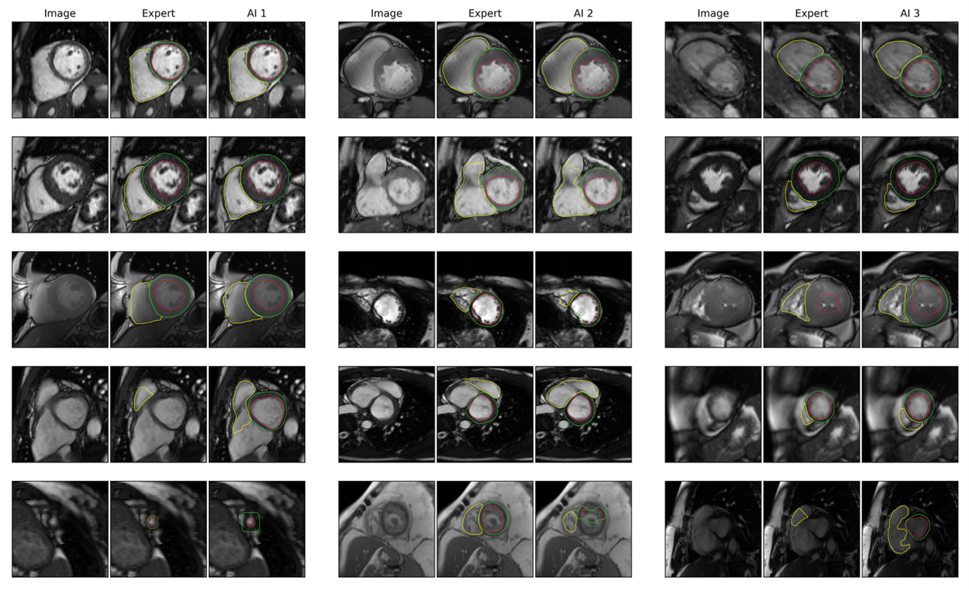
 Figure 2: Segmentation Examples Caption: Each three-column subsection shows the base image (first column), the expert and the AI segmentations (columns 2 & 3). Example images show excellent segmentations in the first two rows. But they also include difficult cases, such as artefacts (section 1, row 3), a large apical thrombus (section 2, row 5) and a hypertrophic heart in end-systole (section 3, row 3).