
Early Career Award Session
Virtual Recording

Xinheng Zhang, PhD
Post-doctoral Scientist
Cedars-Sinai Medical Center
Redwood City, California, United States
Xinheng Zhang, PhD
Post-doctoral Scientist
Cedars-Sinai Medical Center
Redwood City, California, United States
.jpeg "Hsin-Jung (Randy) Yang, PhD photo")
Hsin-Jung (Randy) Yang, PhD
Associate Professor
Biomedical Imaging Research Institute, Cedars-Sinai Medical Center, Los Angeles, CA, USA
Los Angeles, California, United States
Yuheng Huang
Ph. D. Candidate
UCLA, United States
Ghazal Youseff
Graduate student
Indiana University School of Medicine, United States

Anthony G. Christodoulou
Associate Professor
University of California, Los Angeles (UCLA)
Los Angeles, California, United States

Debiao Li, PhD
Professor
Cedars Sinai Medical Center
Los Angeles, California, United States
Andreas Kumar, MD
doctor
Northern Ontario School of Medicine, Sudbury,Ontario, Canada
Sudbury, Ontario, Canada

Rohan Dharmakumar, PhD
Executive Director
Indiana University School of Medicine
Indianapolis, Indiana, United States
 Figure 2. MI Size and Transmurality with Proposed and Conventional Approaches. A) Representative images from canine acquired using conventional and the proposed approaches. D) ROC analysis for MI detection for subjects with acute and chronic timepoints, along with corresponding AUC, are shown. C) – D) Linear regression analysis and bland-altman analysis of MI size and MI transmurality determined using the Conventional and the proposed approaches, across all animals (along with line and equation of best fit, R2 and P values) and corresponding Bland-Altman analysis (along with RPC) are shown.
 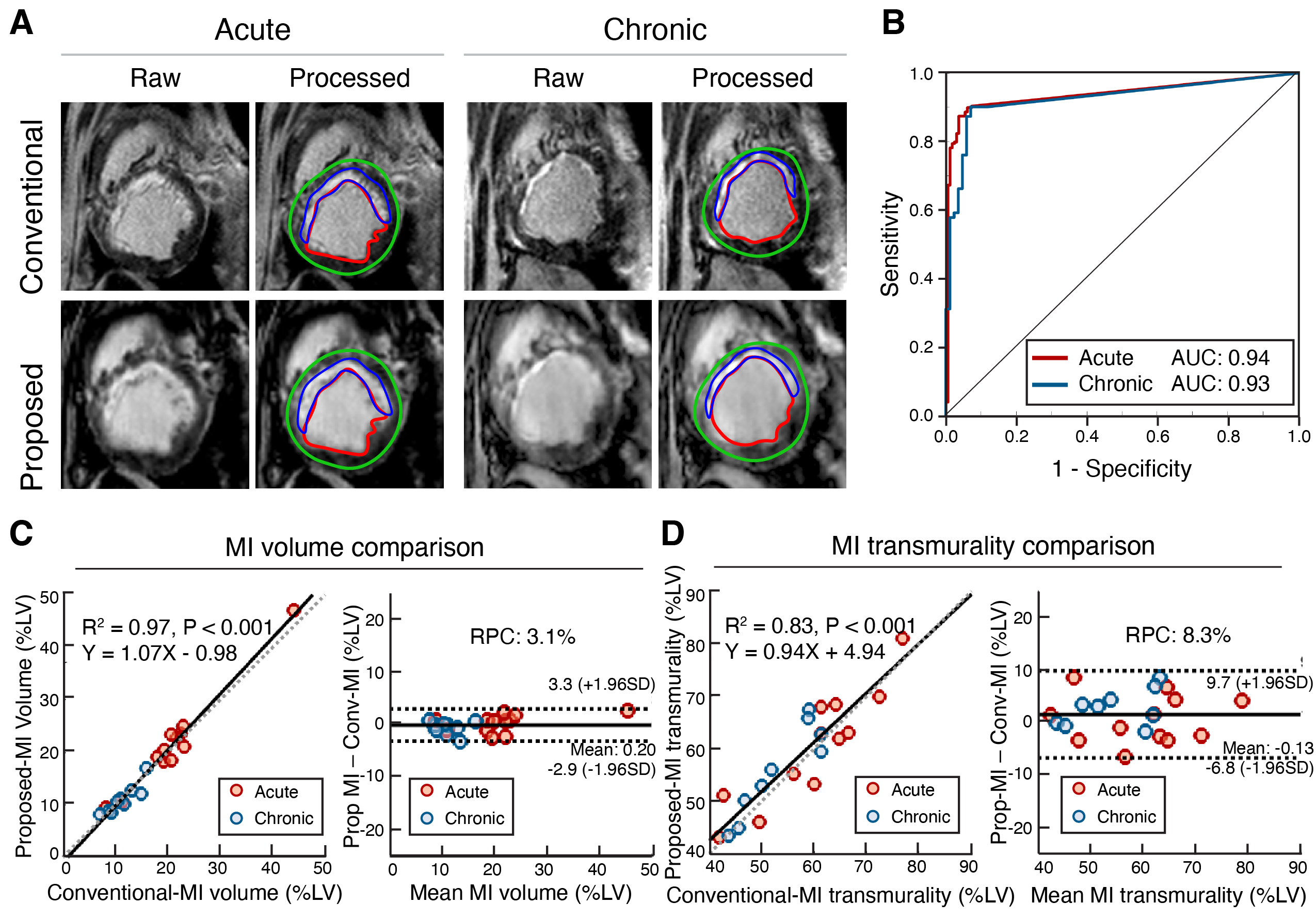 Figure 3. Validation between conventional 2D (Conv.) and proposed (Prop.) measures of microvascular obstruction (MVO) and intramyocardial hemorrhage (IMH): A) and D) are representative subjects of MVO characterization and IMH characterization between the two methods, respectively. B) and E) are linear regression between the two methods in MVO and IMH characterization. C) and F) ROC analysis between the two approaches, the area-under-the-curve (AUC) of ROC analysis is shown. G) Histological validation of MI and IMH using Trichrome and Prussian Blue staining.
.png)
