Quick Fire Session

Farah Cadour, MD, PhD
Assistant professor of cardiothoracic imaging
University Medical Imaging Toronto
Toronto, Ontario, Canada
Farah Cadour, MD, PhD
Assistant professor of cardiothoracic imaging
University Medical Imaging Toronto
Toronto, Ontario, Canada
Felipe Castillo Aravena, MD
Radiologist
University of Toronto, Canada
Jônatas Fávero Prietto dos Santos, MD
Radiologist
Hospital Moinhos de Vento, Brazil
Gabriela Schneider Galvao, MD
Radiologist
Hospital Moinhos de Vento, Brazil
 Figure 2. CT and cardiac MRI correlation showing aortic wall thickening on initial CT (A) and Haste black-blood (C) with resolution of the aortitis on follow-up CT (B) and MRI Haste black-blood (D) after treatment introduction.
 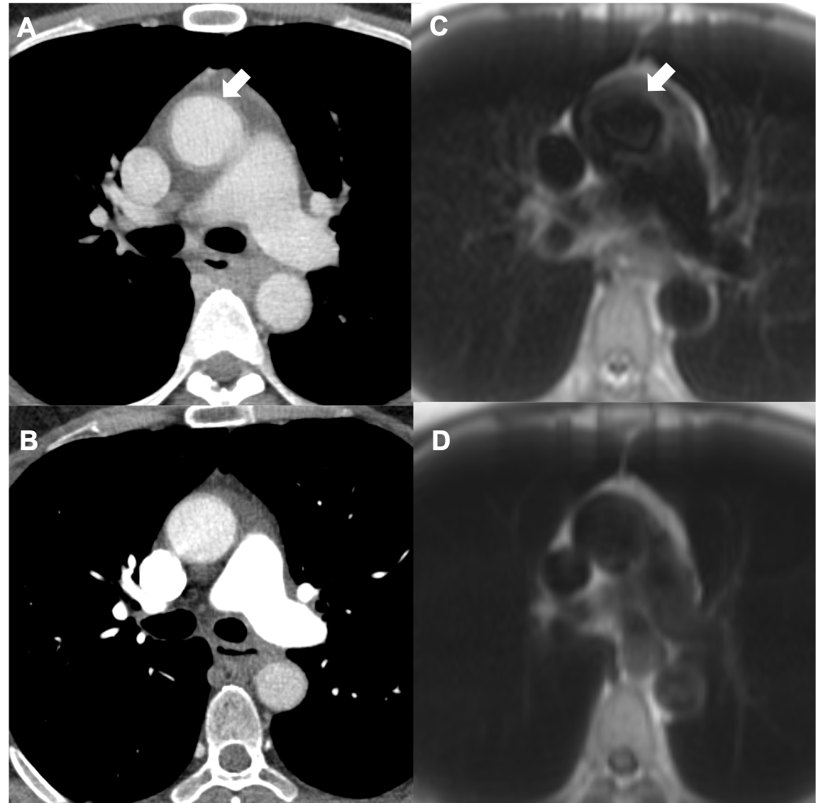 Figure 3. CMR short-axis Turboflash free-breathing PSIR with motion correction shows circumferential subendocardial LGE in the mid (A) to apical (B) left ventricle myocardium and enhancement of the posteromedial papillary muscle (yellow arrow, A), that have mildly improved under treatment in the mid ventricle except for papillary muscle (C) but remains relatively stable at the apex (D).
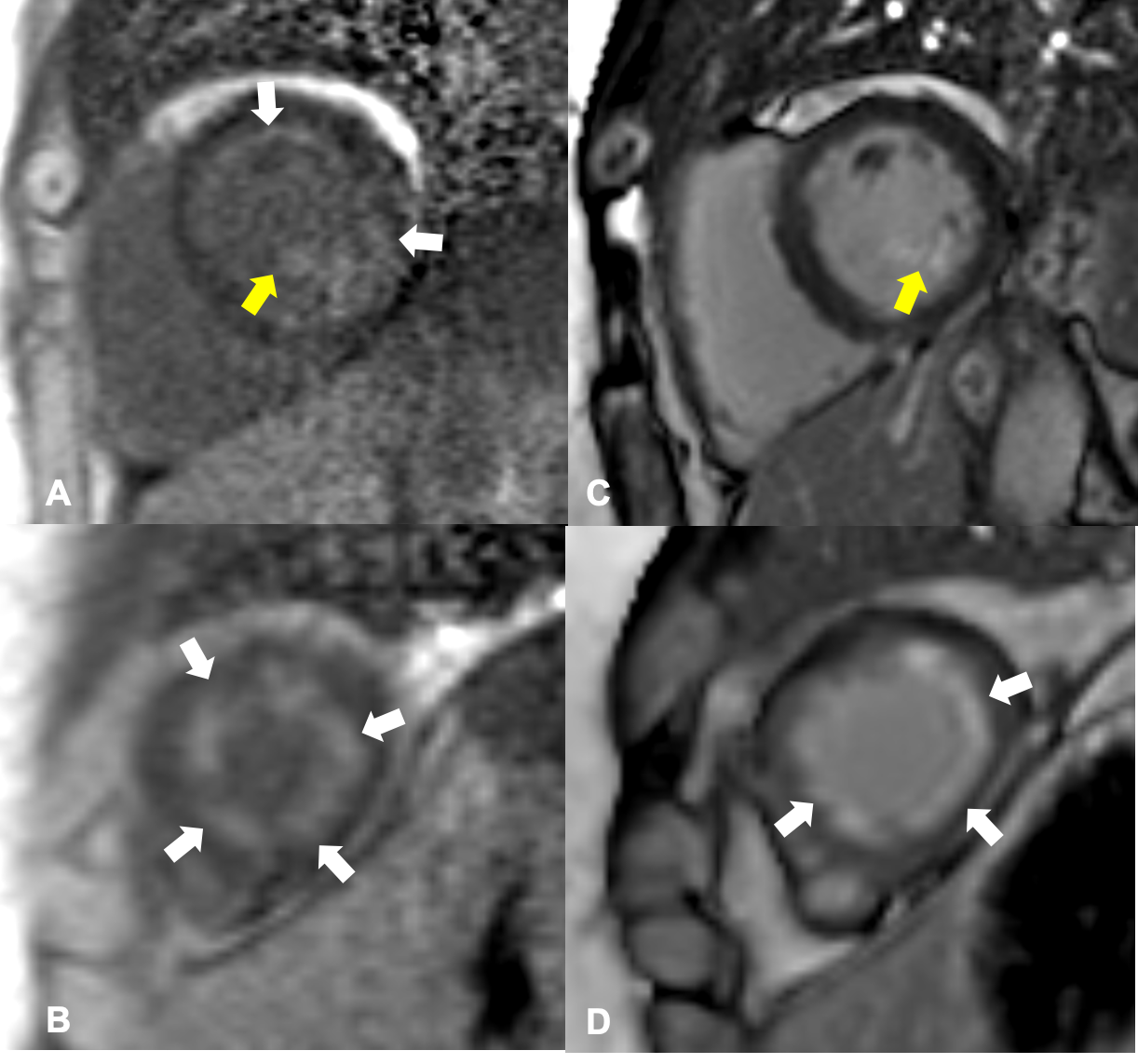