Rapid Fire Session

Stanislas Rapacchi, PhD
Senior researcher
CHUV-UNIL
Lausanne, Vaud, Switzerland
Stanislas Rapacchi, PhD
Senior researcher
CHUV-UNIL
Lausanne, Vaud, Switzerland
Martin Nicoletti, MSc
PhD student
Lausanne University Hospital (CHUV) and University of Lausanne (UNIL)
Lausanne, Vaud, Switzerland
Augustin C. Ogier, PhD
Postdoctoral fellow
University Hospital (CHUV) and University of Lausanne (UNIL), Switzerland
Adèle Mackowiak, PhD
Research associate
University Hospital (CHUV) and University of Lausanne (UNIL)
Lausanne, Switzerland
Jean-Baptiste Ledoux, MSc
Senior Technologist
Lausanne University Hospital (CHUV)
Lausanne, Vaud, Switzerland
Isabel Montón Quesada, MSc
PhD student
Lausanne University Hospital (CHUV)
Lausanne, Vaud, Switzerland

Christopher W. Roy, PhD
Lecturer
University Hospital (CHUV) and University of Lausanne (UNIL)
Lausanne, Vaud, Switzerland

Jérôme Yerly, PhD
Senior scientist
Lausanne University Hospital (CHUV) and University of Lausanne (UNIL)
Lausanne, Vaud, Switzerland
Matthias Stuber, PhD
Professor/Director
CIBM/CHUV/UNIL
Lausanne, Switzerland
full-line FRF | bStar FRF | bNovae FRF | |
Acquisition Parameters | |||
TE/TR (ms) | 2.7 / 5.4 | 0.4, 2.8 / 3.3 | 0.4, 3.7 / 5.1 |
Flip angle (°) | 80 | ||
FOV (mm³) | 300 × 300 × 300 | ||
Spatial resolution (mm³) | 1.5 × 1.5 × 1.5 | ||
Sampling Strategy | |||
Number of segments | 25 | ||
Number of interleaves | 2,812 | 4,601 | 2,978 |
Total acquisition time (min) | 6:00 | ||
Reconstructed respiratory bins × cardiac phases | 4 × 20 | ||
Performance Metrics | |||
Sampling duty cycle† (%) | 44 | 73 | 64 |
Blood-myocardium contrast ratio‡ (%) | 90 ± 31 | 31 ± 18 | 72 ± 19 |
Image sharpness (a.u.) | 0.61 ± 0.29 | 2.68 ± 3.40 | 1.12 ± 1.95 |
Abbreviations: FRF, free-running framework; FOV, field of view; TE, echo time; TR, repetition time; a.u., arbitrary units.
Notes: † Sampling duty cycle calculated as ADC duration/TR × 100% ‡ Blood-myocardium contrast ratio calculated as (B-M)/M × 100%, where B represents blood signal intensity and M represents myocardial signal intensity
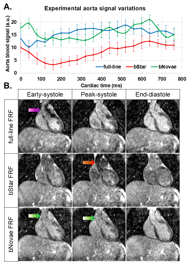
Free-running sequence modules (A), with ADC sampling in gray, were included in a numerical simulation (B) to confirm bStar high flow sensitivity (C). By extending the 2nd echo, in proposed bNovae, flow-induced signal loss was greatly reduced while maintaining an ultra-short echo time and a high sampling duty cycle..png) A: Measurements of free-running (FRF) signal variations in the aortic root confirmed bStar signal loss, as anticipated by numerical simulations. B: Coronal views of key frames within the cardiac cycle showed severe signal loss in the aortic root: from intravoxel dephasing in full-line FRF (purple arrow) and from flow-induced accumulated phase in bStar FRF (red arrow). Thanks to increased sampling efficiency, bStar and bNovae images were also sharper than full-line images that suffered from a global mild blur.
A: Measurements of free-running (FRF) signal variations in the aortic root confirmed bStar signal loss, as anticipated by numerical simulations. B: Coronal views of key frames within the cardiac cycle showed severe signal loss in the aortic root: from intravoxel dephasing in full-line FRF (purple arrow) and from flow-induced accumulated phase in bStar FRF (red arrow). Thanks to increased sampling efficiency, bStar and bNovae images were also sharper than full-line images that suffered from a global mild blur.