Rapid Fire Session

Mohammad Q. Mehdi, MD
Pediatric Advanced Cardiac Imaging Fellow
The University of Texas Southwestern Medical Center
Dallas, Texas, United States
Mohammad Q. Mehdi, MD
Pediatric Advanced Cardiac Imaging Fellow
The University of Texas Southwestern Medical Center
Dallas, Texas, United States

Fares Munes, MD
Assistant Professor
Atrium Health Levine Children’s Hospital / Wake Forest School of Medicine
Charlotte, North Carolina, United States
Yuland Tyner, BA, RT
Cardiac MRI Technologist
Children's Hospital Dallas
Lewisville, Texas, United States
Steven Philip, RT
MRI Technologist
Childrens's Medical Center Dallas
Dallas, Texas, United States
Buford S. Scott, RT
Senior Pediatric Cardiac Technologist
Childrens Health Systems
Dallas, Texas, United States
Jennifer Smith, MD
Pediatric Radiologist
The University of Texas Southwestern Medical Center, United States

Mansi Gaitonde, MD
Assistant Professor
The University of Texas Southwestern Medical Center
Dallas, Texas, United States
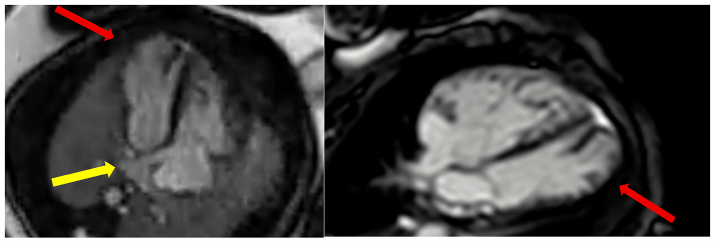
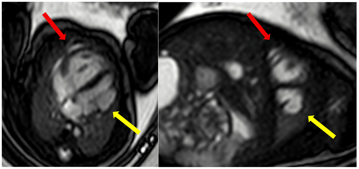 Figure 2: Fetal cardiac magnetic resonance (CMR) imaging in a 35 week fetus. Fetal Doppler-derived ECG gated balanced steady state free procession cine imaging in 4-chamber plane (left panel) shows a dilated coronary sinus with anomalous pulmonary venous drainage of at least three pulmonary veins draining into the coronary sinus (yellow arrow). Additionally, a large mid to apical left ventricular diverticulum was identified measuring 25 × 9 mm in diastole and 17 × 7 mm in systole (red arrow). Cine imaging on postnatal CMR (right panel) done in the first week of life with a comparative 4 chamber plane showing the left ventricular diverticulum (red arrow).