Oral Abstracts Session
Virtual Recording

Dabne Celin Barrera Trujillo, MSc
Master Student
Pontificia Universidad Católica de Chile
Santiago, Region Metropolitana, Chile

Claudia Prieto, PhD
Professor
Pontificia Universidad Católica de Chile / King's College London
Santiago, Region Metropolitana, Chile
Rafael De la sotta, MSc
Pre-doctoral engineer
Millennium Institute for Intelligent Healthcare Engineering, Chile

Rene M. Botnar
Professor of Cardiovascular Imaging
Institute for Biological and Medical Engineering, Pontificia Universidad Católica de Chile
Santiago de Chile, Region Metropolitana, Chile
.jpg "Karl P. Kunze, PhD photo")
Karl P. Kunze, PhD
Senior Cardiac MR Scientist
Siemens Healthineers
Camberley, England, United Kingdom
Carlos Castillo-Passi, PhD
Post-doctoral stundent
Stanford University, United States
.png) Figure 2. (a). Phantom results for 3D MUST-T2 at 0.55T. Plots compare the mean T2 values derived from the 9 vials for MUST-T2 and T2p-bSSFP with the ground-truth T2 values (measured by spin echo [SE] with 8 TEs from 10-640 ms). (b). T2 maps obtained using the free-breathing 3D MUST-T2 and the conventional breath-held 2D T2p-SSFP sequences at 0.55T for 3 healthy subjects. The 3D MUST-T2 slices were reformatted to short axis to match the 2D T2 map acquisitions. Good visualization of the myocardium and surrounding structures can be observed on the 3D MUST-T2 maps. Acquisition times are expressed as minutes: seconds. Abbreviations: AT, acquisition time.
Figure 2. (a). Phantom results for 3D MUST-T2 at 0.55T. Plots compare the mean T2 values derived from the 9 vials for MUST-T2 and T2p-bSSFP with the ground-truth T2 values (measured by spin echo [SE] with 8 TEs from 10-640 ms). (b). T2 maps obtained using the free-breathing 3D MUST-T2 and the conventional breath-held 2D T2p-SSFP sequences at 0.55T for 3 healthy subjects. The 3D MUST-T2 slices were reformatted to short axis to match the 2D T2 map acquisitions. Good visualization of the myocardium and surrounding structures can be observed on the 3D MUST-T2 maps. Acquisition times are expressed as minutes: seconds. Abbreviations: AT, acquisition time..png) Figure 3. The T2 accuracy of the 3D MUST-T2 sequence at 0.55T versus conventional 2D T2p-SSFP, as measured by the mean T2 value, are shown in the left ventricular segmentation. The T2 values measured with 3D MUST-T2 are in good agreement with the literature (T2 = 48.7 ± 4.3 ms), showing underestimation with respect to 2D T2p-SSFP (3).
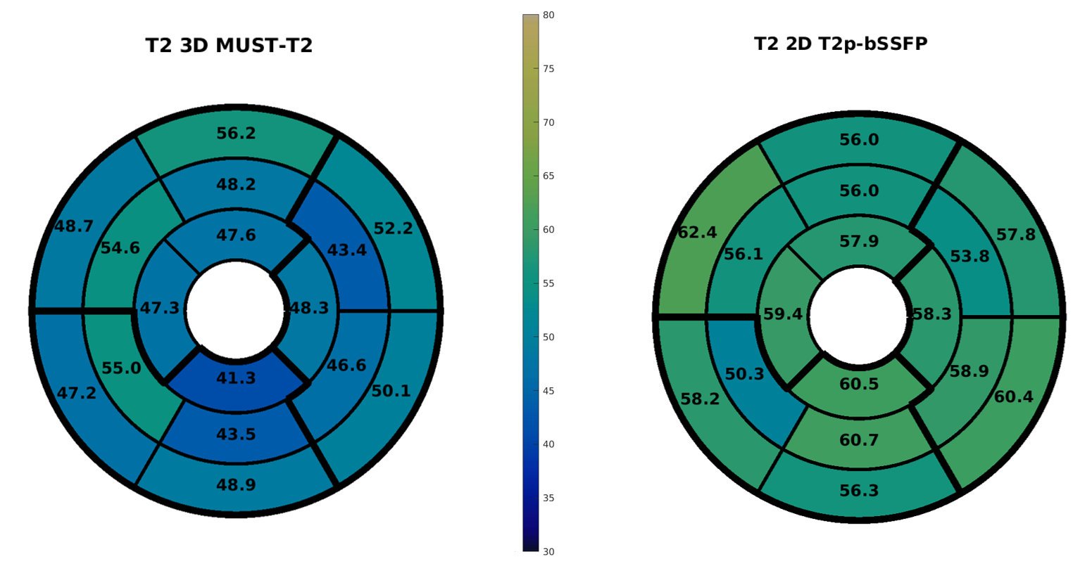
Figure 3. The T2 accuracy of the 3D MUST-T2 sequence at 0.55T versus conventional 2D T2p-SSFP, as measured by the mean T2 value, are shown in the left ventricular segmentation. The T2 values measured with 3D MUST-T2 are in good agreement with the literature (T2 = 48.7 ± 4.3 ms), showing underestimation with respect to 2D T2p-SSFP (3).