Case Session
Virtual Recording

Parker Rushworth, MD
Advanced Cardiovascular Imaging Fellow
University of California, San Francisco
Emeryville, California, United States
Parker Rushworth, MD
Advanced Cardiovascular Imaging Fellow
University of California, San Francisco
Emeryville, California, United States
Dany Cheikh-Debs, MD
Advanced Cardiovascular Imaging Fellow
University of California, San Francisco, United States

Christopher Lee, MD
Assistant Professor
University of California, San Francisco
San Francisco, California, United States

Michael Salerno, MD, PhD
Professor of Medicine and Radiology and Biomedical Imaging
UCSF
Belmont, California, United States
.jpg) Fig 2. SSFP cine short axis images in end diastole demonstrating significant LV hypertrophy of the (A) (B) LV base, (C) LV mid cavity, and (D) (E) LV apex. The second row of images demonstrate short axis views with the presence of diffuse late gadolinium in the (F) (G) LV base, (H) LV mid cavity, and (I) (J) LV apex.
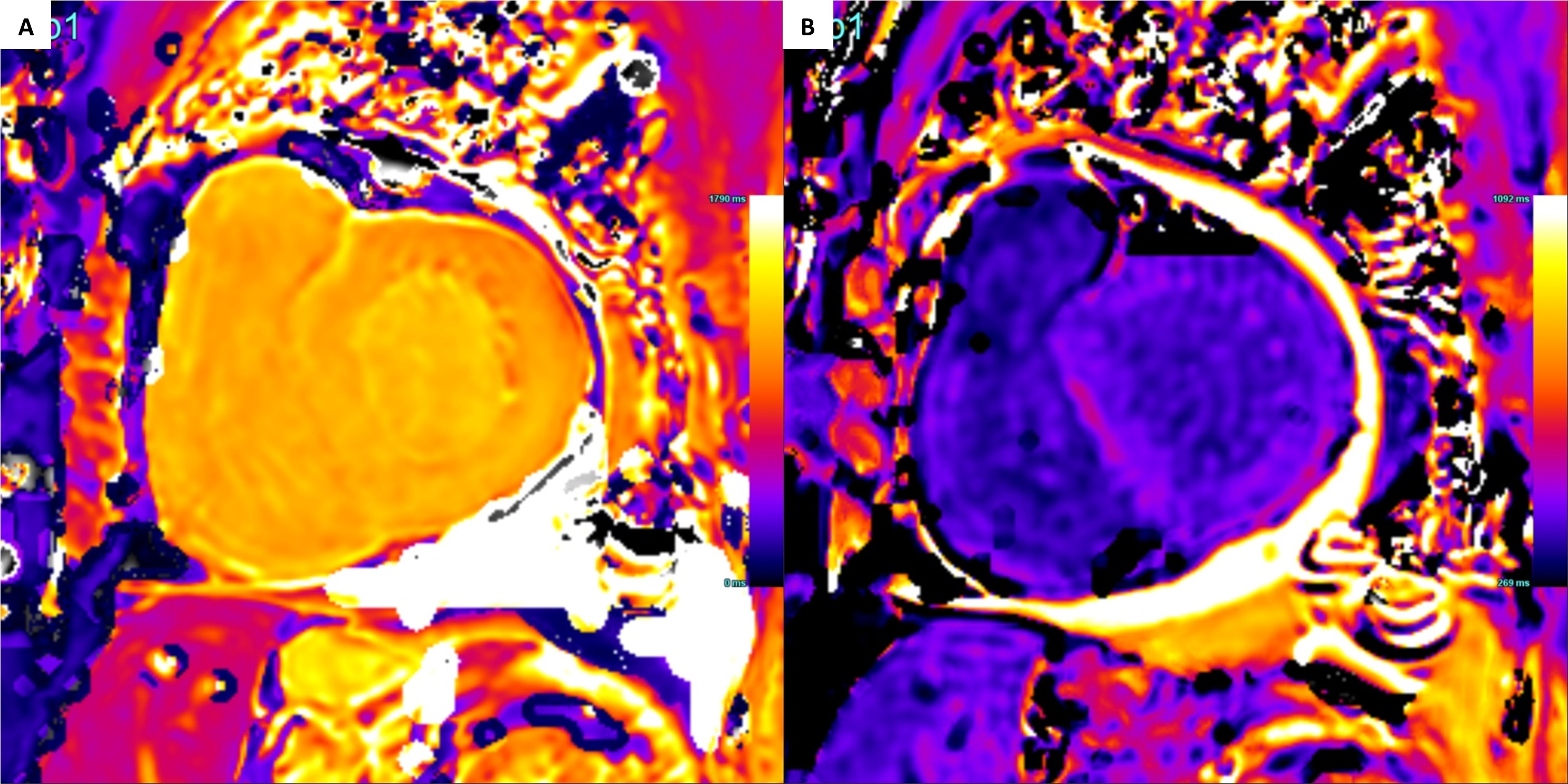
Fig 2. SSFP cine short axis images in end diastole demonstrating significant LV hypertrophy of the (A) (B) LV base, (C) LV mid cavity, and (D) (E) LV apex. The second row of images demonstrate short axis views with the presence of diffuse late gadolinium in the (F) (G) LV base, (H) LV mid cavity, and (I) (J) LV apex.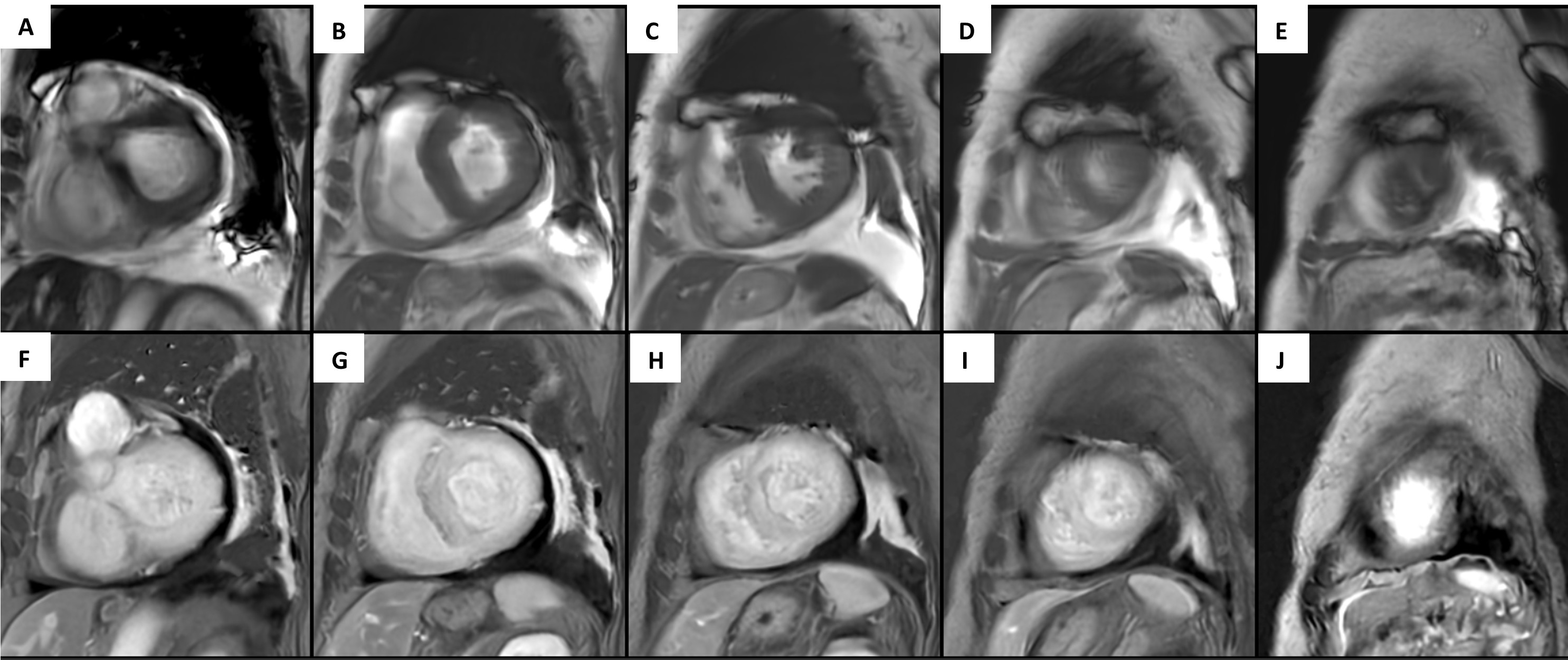 Fig 3. (A) Native T1 mapping of the LV base demonstrating a normal myocardial value of 1132ms and an unexpectedly low blood pool value of 1237ms (Acquired on a 3T machine). (B) Post-contrast T1 mapping of the LV base demonstrating a normal myocardial value of 433ms and blood pool value of 406ms. These myocardial values are inappropriately low for a patient with biopsy confirmed AL amyloidosis given concurrent use of supplemental IV iron.