Quick Fire Session

Gabriel Amaral Mendes Ferreira, MD
Cardiologist
Universidade Federal de São Paulo - Unifesp/ DASA
Sao Paulo, Sao Paulo, Brazil
Gabriel Amaral Mendes Ferreira, MD
Cardiologist
Universidade Federal de São Paulo - Unifesp/ DASA
Sao Paulo, Sao Paulo, Brazil
Guilherme Martins Guzman
Cardiologistgis
Hospital 9 de Julho, Brazil
Marcia de Paula Cardinali
Cardiologistgist
DIAGNÓSTICOS DA AMÉRICA S.A., Brazil

Maira Azevedo Ximenes
Fellowship in Cardiac Imaging Department DASA + Unifesp
Universidade Federal de São Paulo - Unifesp
São Paulo, Sao Paulo, Brazil

Marly Uellendahl, MD, PhD
Cardiac Imaging Coordinator
DASA - UNIFESP
São Paulo, Sao Paulo, Brazil
Alfredo Eyer Rodrigues
Cardiologist
Universidade Federal de São Paulo - Unifesp, Brazil
Cecilia Maria Pereira Pereira Santos
Fellowship in Cardiac Imaging DASA-SP
Universidade Federal de São Paulo - Unifesp
São Paulo, Sao Paulo, Brazil
 Image 2: Cardiac magnetic resonance. Left: tubular structure in the SVC with susceptibility artifact extending into the RA (white arrow). Right: single-shot LGE PSIR demonstrating contrast opacification within the stent lumen, consistent with preserved flow (yellow arrow).
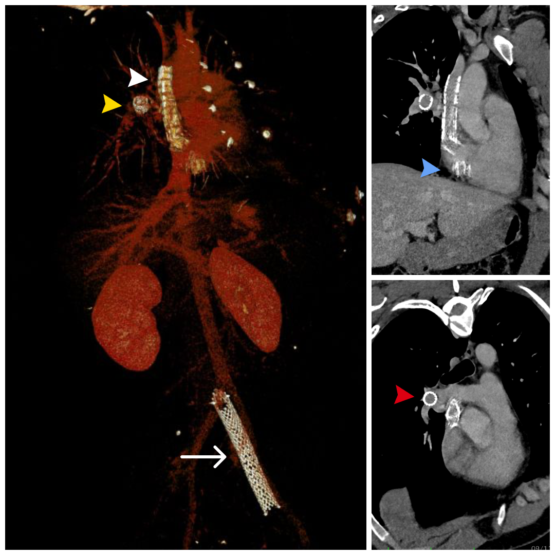
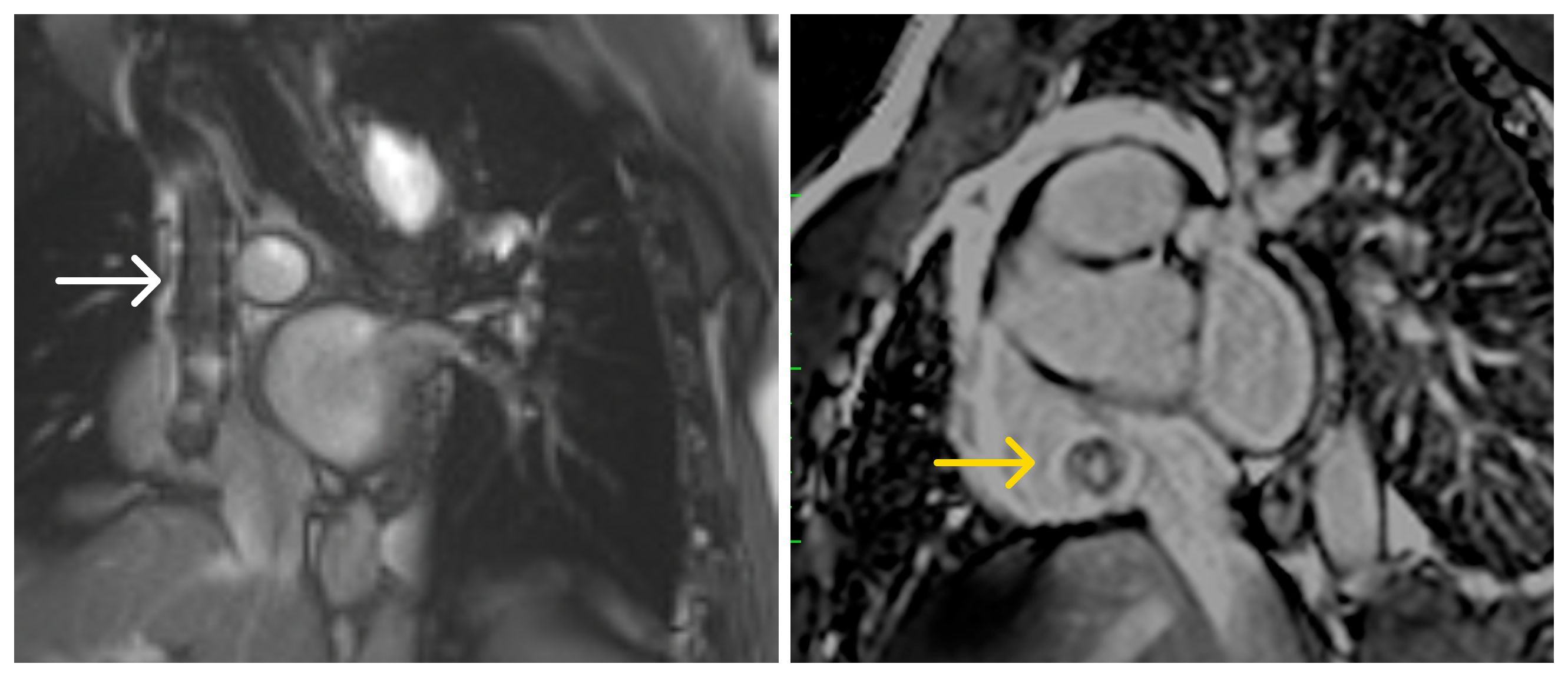 Image 3: Computed tomography. Left: 3D CTA reconstruction showing a stent in the left common iliac vein (white arrow), the embolized stent positioned within the RA with a fractured proximal edge and with its distal end lodged in the SVC (white arrowhead) and a small stent fragment located in the right pulmonary artery branch (yellow arrowhead). Upper right: View highlighting the relationship of the stent between the SVC and RA, with evidence of a fractured distal edge (blue arrowhead). Lower right: Axial view at the level of the pulmonary artery showing a metallic fragment in the right pulmonary artery branch (red arrowhead).