Quick Fire Session

David Martínez Juárez, MD
Radiology
Christus muguerza
Puebla, Puebla, Mexico
David Martínez Juárez, MD
Radiology
Christus muguerza
Puebla, Puebla, Mexico
Francisco Zamora Rosales
chief investigator
christus Muguerza, Puebla, Mexico
omar Gomez Monterrosas
cardiologist
christus Muguerza, Puebla, Mexico
 Figure 2: Cardiac magnetic resonance imaging. Image A-C: LGE sequences in short-axis (A and B) and four-chamber (C) views showing subepicardial enhancement in the posterior and inferior walls (yellow arrows), a pattern consistent with non-ischemic myocardial injury of probable inflammatory origin. Image D: First-pass perfusion sequence demonstrating a subendocardial perfusion defect in the septal and inferior walls of the mid-apical segment (yellow arrows), suggestive of ischemia without a clear coronary territory.
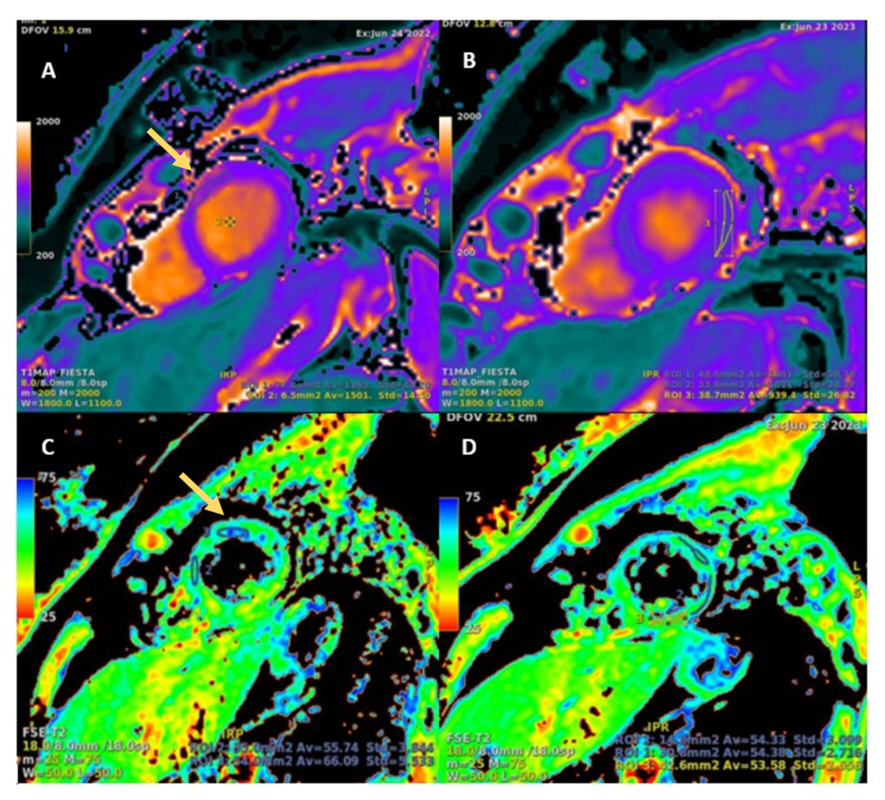
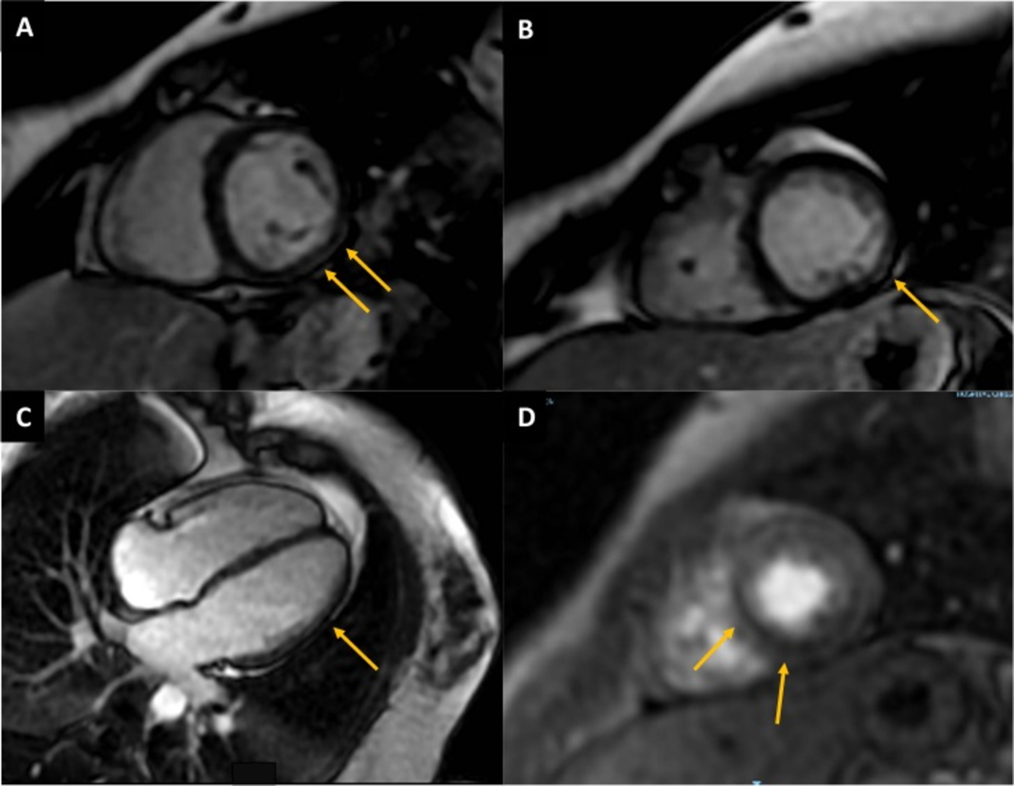 Figure 3: CMR T1 native and T2 mapping. Baseline and Follow-up study one year later. Image A and C: Short-axis view of the left ventricle mid segment. Baseline study showing increased relaxation times with focal elevation of native T1 mapping in the anterior wall (1153 ms, yellow arrows) and T2 mapping of 60 ms. Image B and D: Follow-up study performed one year later demonstrates a reduction in anterior wall relaxation times (native T1 mapping 1021 ms, T2 mapping 54 ms).