Rapid Fire Session
Rebecka Steffen Johansson, MD
MD, PhD student
Karolinska Institutet
Stockholm, Stockholms Lan, Sweden
Rebecka Steffen Johansson, MD
MD, PhD student
Karolinska Institutet
Stockholm, Stockholms Lan, Sweden
Christina Ekenbäck, MD
Karolinska Institutet Danderyds Sjukhus, Sweden
Elin B. Brolin, MD, PhD
Karolinska Institutet, Sweden
Jens Jensen, MD, PhD
MD, Associate Professor
Karolinska Institute, Sweden
Martin G. Sundqvist, MD, PhD
MD, PhD
Karolinska Institute, Sweden
Shams Y-Hassan, MD
MD
Karolinska Institute, Sweden
Loghman Henareh, MD, PhD
MD, Associate Professor
Karolinska Institute, Sweden
Kenneth Caidahl, MD, PhD
Professor
Karolinska Institutet
Stockholm, Stockholms Lan, Sweden
Jonas Spaak, MD, PhD
MD, Professor
Karolinska Institutet
Stockholm, Stockholms Lan, Sweden
Andreas Sigfridsson, PhD
Associate Professor
Karolinska Institute, Sweden
Peter Kellman, PhD
Dr.
National Institutes of Health, Maryland, United States
.jpg "Martin Ugander, MD, PhD photo")
Martin Ugander, MD, PhD
Professor of Cardiac Imaging
Kolling Institute, Royal North Shore Hospital and University of Sydney/ Karolinska University Hospital and Karolinska Institutet
Sydney, New South Wales, Australia
Per Tornvall, MD, PhD
Karolinska Institute, Stockholms Lan, Sweden
Peder Sörensson, MD, PhD
Associate professor
Karolinska Institute, Stockholms Lan, Sweden

Jannike Nickander, MD, PhD
MD, PhD
Karolinska Institutet
Stockholm, Stockholms Lan, Sweden
Clinical characteristics | Index (n=42) | Follow-up (n=42) | Change, p</em> |
Age (years) | 60 [55-66] | 61 [55-66] | < 0.001 |
Height (cm) | 167 [162-172] | 167 [161-171] | 0.38 |
Weight (kg) | 70 [62-80] | 70 [61-80] | 0.65 |
BSA (m2) | 1.8 [1.7-1.9] | 1.8 [1.7-1.9] | 0.81 |
Initial Troponin T (ng/L) | 55 [36-126] |
|
|
Maximal Troponin T (ng/L) | 254 [115-438] |
|
|
NT-proBNP (ng/L) | 795 [437-1670] |
|
|
Hypertension | 9 (21%) |
|
|
Hyperlipidemia | 6 (14%) |
|
|
Diabetes mellitus | 3 (7%) |
|
|
Smoking currently | 8 (19%) |
|
|
Smoking previously | 14 (33%) |
|
|
TTS ballooning pattern on initial CMR | |||
Midventricular | 32 (76%) |
|
|
Mid-lateral | 2 (5%) |
|
|
Midventricular and apical | 1 (2%) |
|
|
Apical | 7 (17%) |
|
|
CMR findings | |||
LVEDV (ml) | 146 [131-155] | 139 [126-156] | Δ -5%, p=0.80 |
LVEDVi (ml/m2) | 81 [73-88] | 81 [73-86] | Δ 0%, p=0.70 |
LVESV (ml) | 65 [50-73] | 59 [51-67] | Δ -9%, p=0.008 |
LVESVi (ml/m2) | 35 [32-42] | 33 [28-38] | Δ -6%, p=0.005 |
LVSV (ml) | 79 [68-87] | 81 [75-94] | Δ 3%, p=0.002† |
LVSVi (ml/m2) | 44 [40-49] | 47 [43-52] | Δ 7%, p=0.002† |
LVEF (%) | 55 [51-58] | 59 [55-63] | Δ 7%, p< 0.001 |
LVM (g) | 112 [103-133] | 110 [101-121] | Δ -2%, p=0.22 |
LVMi (g/m2) | 64 [58-71] | 63 [57-67] | Δ -2%, p=0.18 |
Native T1 (ms) | 1102 [1072-1142] | 1015 [995-1040] | Δ -8%, p< 0.001† |
ECV (%) | 31 [30-34] | 28 [26-30] | Δ -10%, p< 0.001 |
GLS (%) | -16 [-17-(-13)] | -20 [-20-(-18)] | Δ 25%, p< 0.001 |
GCS (%) | -14 [-17-(-11)] | -20 [-22-(-18)] | Δ 43%, p< 0.001 |
Data presented as median [interquartile range] or n (%), change denoted Δ represents median percent change, p-values denotes the Wilcoxon signed-rank test and p-values marked with † denotes the paired t-test. Abbreviations: CMR = cardiovascular magnetic resonance imaging; BSA = body surface area; LVEDV = left ventricular end diastolic volume; LVESV = left ventricular end systolic volume; LVSV = left ventricular stroke volume; LVEF = ejection fraction, LVM = left ventricular mass, ECV = extracellular volume; GLS = global longitudinal strain; GCS = global circumferential strain; NT-proBNP = N-terminal pro–B-type natriuretic peptide; i signifies indexed to BSA.
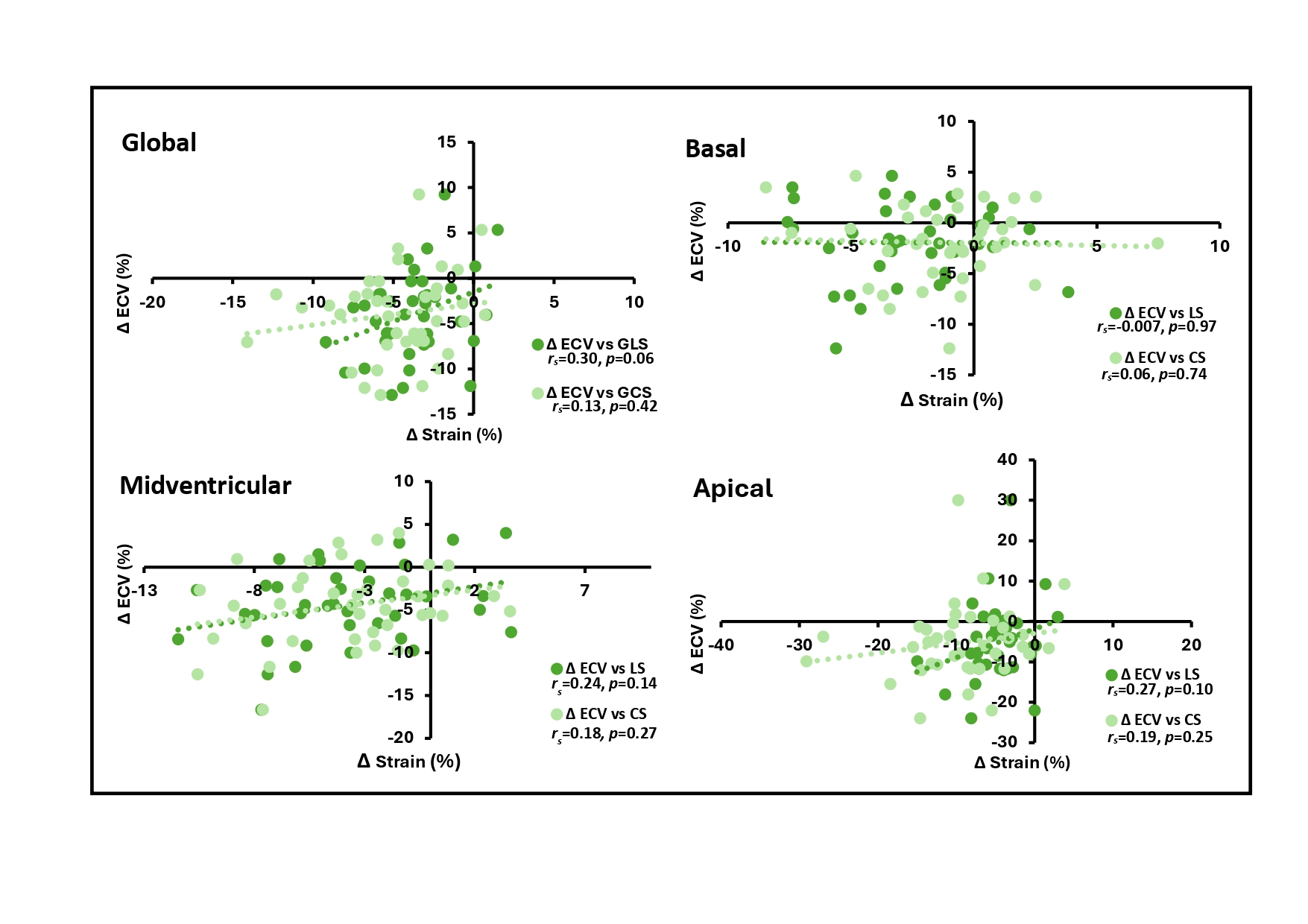
Figure 1. Scatterplots of native T1 vs LS and CS. The scatterplots show change of global, basal, midventricular, and apical native T1 vs LS and CS, with Spearman’s rank correlation coefficient rs and p-values. Abbreviations: LS = longitudinal strain; CS = circumferential strain.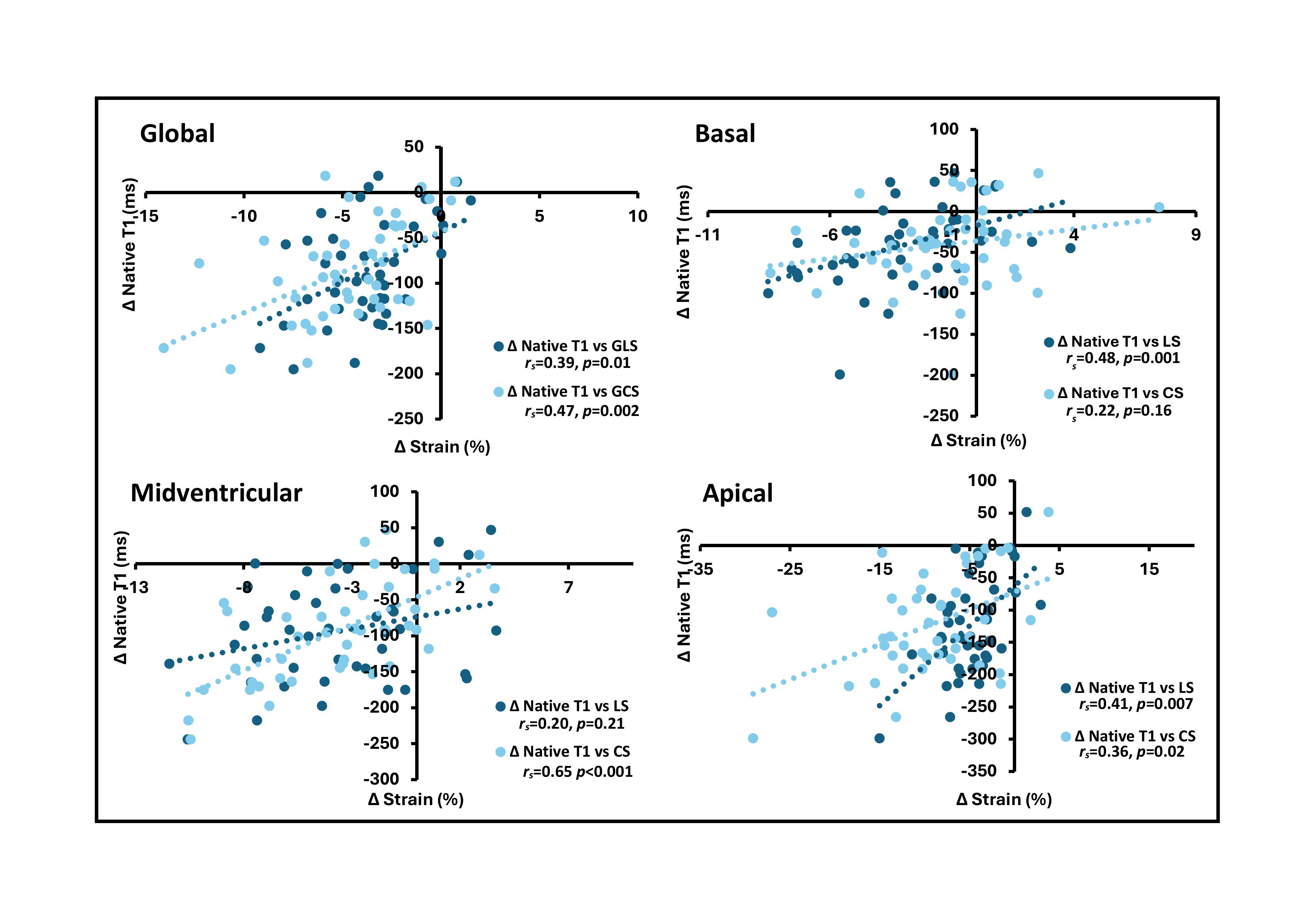 Figure 2. Scatterplots of ECV vs LS and CS. The scatterplots show change of global, basal, midventricular, and apical ECV vs LS and CS, with Spearman’s rank correlation coefficient rs and p-values. Abbreviations: ECV = extracellular volume; LS = longitudinal strain; CS = circumferential strain.