Rapid Fire Session

Augustin C. Ogier, PhD
Post-doctoral fellow
Lausanne University Hospital (CHUV) and University of Lausanne (UNIL)
Lausanne, Vaud, Switzerland
Augustin C. Ogier, PhD
Post-doctoral fellow
Lausanne University Hospital (CHUV) and University of Lausanne (UNIL)
Lausanne, Vaud, Switzerland
Gabriel Paffi, MSc
MSc student
University Hospital (CHUV) and University of Lausanne (UNIL), Switzerland

Jérôme Yerly, PhD
Senior scientist
Lausanne University Hospital (CHUV) and University of Lausanne (UNIL)
Lausanne, Vaud, Switzerland
Angela Rocca, MSc
PhD student
Lausanne University Hospital (CHUV) and University of Lausanne (UNIL)
Lausanne, Vaud, Switzerland
Isabel Montón Quesada, MSc
PhD student
Lausanne University Hospital (CHUV)
Lausanne, Vaud, Switzerland
Jean-Baptiste Ledoux, MSc
Senior Technologist
Lausanne University Hospital (CHUV)
Lausanne, Vaud, Switzerland

Robert J. Holtackers, PhD
MR Physicist & Assistant Professor
Maastricht University Medical Centre
Maastricht, Limburg, Netherlands
Matthias Stuber, PhD
Professor/Director
CIBM/CHUV/UNIL
Lausanne, Switzerland

Christopher W. Roy, PhD
Lecturer
University Hospital (CHUV) and University of Lausanne (UNIL)
Lausanne, Vaud, Switzerland
Roger Hullin, MD
Chief Severe Heart Failure en Heart Transplantation Program
Lausanne University Hospital (CHUV)
Lausanne, Vaud, Switzerland
David Rotzinger, MD
Associate radiologist
Lausanne University Hospital (CHUV) and University of Lausanne (UNIL)
Lausanne, Vaud, Switzerland

Ruud B. van Heeswijk, PhD, FSCMR
Senior lecturer
Lausanne University Hospital (CHUV) and University of Lausanne (UNIL)
Lausanne, Vaud, Switzerland
Metric | Region | Model | ||
CS-FRA | fNav-FRA | Gridded-FRA | ||
DSC (1) | LVB | 0.94 ± 0.01 | 0.93 ± 0.02 | 0.93 ± 0.01 |
LVM | 0.86 ± 0.02 | 0.82 ± 0.04 | 0.82 ± 0.04 | |
RVB | 0.92 ± 0.02 | 0.90 ± 0.02 | 0.91 ± 0.02 | |
RVD (%) | LVB | 2.4 ± 2.1 | 3.1 ± 2.4 | 4.1 ± 2.5 |
LVM | 5.8 ± 3.7 | 6.6 ± 4.3 | 7.0 ± 5.3 | |
RVB | 4.5 ± 2.8 | 5.6 ± 4.4 | 5.2 ± 3.8 | |
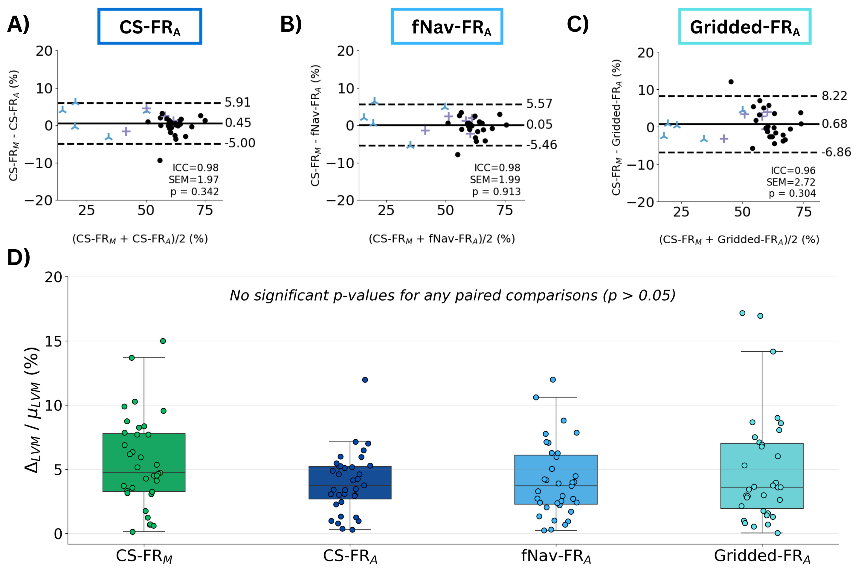
 Figure 2: Bland-Altman analysis of left-ventricular ejection fraction comparing semi-manual segmentation on compressed-sensing images (CS-FRM) with the DL-based automatic segmentations trained and applied to A) compressed-sensing images (CS-FRA), B) respiratory motion corrected images (fNav-FRA) and C) gridded images (Gridded-FRA). D) Systolic–diastolic left ventricular myocardial (LVM) volume mismatch computed for each segmentation as the difference between the end-diastole and end-systole volumes (∆_LVM) divided by the mean volume (μ_LVM).