Rapid Fire Session

Melany B. Atkins, MD
Division Chief, Cardiovascular Imaging
Fairfax Radiological Consultants, Inova Fairfax Hospital
Arlington, Virginia, United States
Melany B. Atkins, MD
Division Chief, Cardiovascular Imaging
Fairfax Radiological Consultants, Inova Fairfax Hospital
Arlington, Virginia, United States
Junjie Ma, PhD
Cardiac MRI Scientist
GE HealthCare
Jersey City, New Jersey, United States
Haonan Wang, PhD
Lead Cardiac MR Scientist
GE HealthCare
Waukesha, Wisconsin, United States
Ke Li, PhD
Senior PSD and Applications Engineer
GE Healthcare, United States
Alessandro Scotti
MR Clinical Scientist
GE Healthcare, United States
Helen Shengyu Lu, RT
MRI technologist
Inova Fairfax Hospital, United States
Santosh Shah, RT
MRI Technologist
Fairfax Radiology
Springfield, Virginia, United States
Asma Sherazi, RT
MRI Technologist
Fairfax Radiology Consultants/ Inova
woodbridge, Virginia, United States
Michael Vinsky, RT
Academic Clinical Excellence Leader
Inova Fairfax Hospital
Evington, Virginia, United States

Martin A. Janich, PhD
Director, Cardiac MRI
GE HealthCare
Munich, Bayern, Germany
| Conventional protocol | Enhanced protocol |
FOV [cm] | 36.0 | |
Phase FOV | 0.9 | 0.9 |
Slice Thickness [mm] | 8.0 | |
Flip angle [°] | 45 | |
Bandwidth [Hz] | 83.33 | |
Acceleration | ARC x 3 | |
NEX | 1 | 0.9 |
TR [ms] | 3.6 | 3.1 |
TE [ms] | 1.5 | 1.1 |
Spatial Res [mm] | 1.8 | 1.8 |
Temp res [ms] | 258 | 203 |
Figure 1. Representative ssh PS LGE images from a 19-year-old male patient acquired during the 6-month follow-up. The patient presented to the emergency department with one week history of cough, fever, and chest pain. His cardiac MR findings were consistent with acute myocarditis including myocardial edema with elevated T2 value, elevated T1/ECV value, and extensive delayed enhancement. Patient returned 6 months later for follow up with resolution of parametric mapping abnormalities and persistent delayed enhancement. Ssh PS LGE images acquired with the enhanced protocol (A, C, E) demonstrate improved image sharpness and decreased image blur compared to those acquired with the conventional protocol (B, D, F), as highlighted by the yellow arrows. 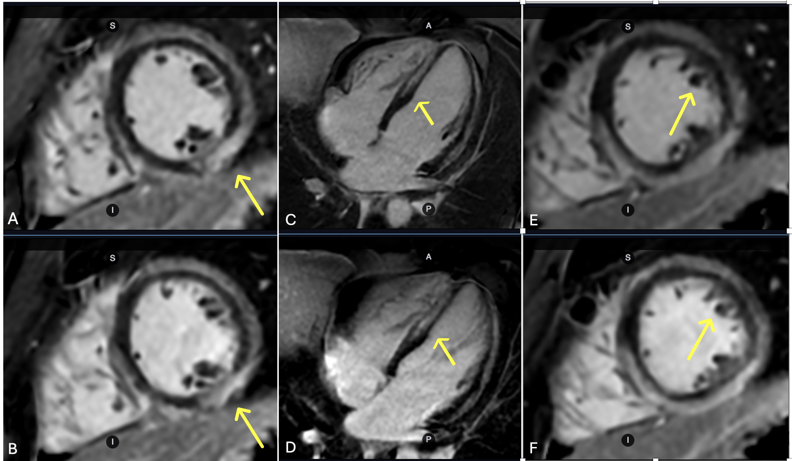 Figure 2. 83-year-old male patient with history of sarcoid referred for evaluation of non-sustained ventricular tachycardia. Ssh PS LGE acquired with the enhanced protocol (A) demonstrates improved image sharpness with better visualization of the inferolateral wall patchy midmyocardial delayed enhancement (yellow arrow) over conventional (C). Additional short axis image demonstrating patchy mid myocardial delayed enhancement within the mid anterolateral wall (white arrow) with improved image sharpness and less image blurring with the enhanced ssh PS LGE (B) over conventional (D).
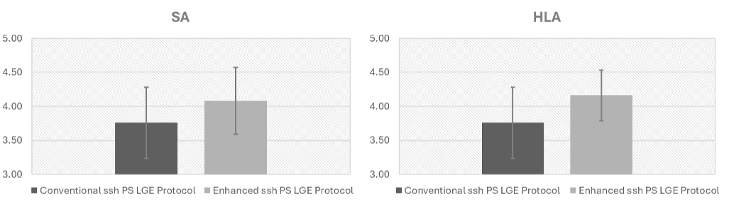
 .png) Figure 3. Comparison of image quality scores for ssh PS LGE acquired using conventional and enhanced protocols (n = 25). On the short-axis (SA) plane (left), the mean score was 4.08 ± 0.49 for the enhanced protocol versus 3.76 ± 0.52 for the conventional protocol (p < 0.05). On the 4-chamber (4CH) plane (right), scores were 4.16 ± 0.37 versus 3.76 ± 0.52, respectively (p < 0.01).
Figure 3. Comparison of image quality scores for ssh PS LGE acquired using conventional and enhanced protocols (n = 25). On the short-axis (SA) plane (left), the mean score was 4.08 ± 0.49 for the enhanced protocol versus 3.76 ± 0.52 for the conventional protocol (p < 0.05). On the 4-chamber (4CH) plane (right), scores were 4.16 ± 0.37 versus 3.76 ± 0.52, respectively (p < 0.01).