Rapid Fire Session

Benjamin Böttcher, MD
Resident / Physician
Institute for Diagnostic and Interventional Radiology, Pediatric Radiology and Neuroradiology / University Medical Center Rostock
Rostock, Mecklenburg-Vorpommern, Germany
Benjamin Böttcher, MD
Resident / Physician
Institute for Diagnostic and Interventional Radiology, Pediatric Radiology and Neuroradiology / University Medical Center Rostock
Rostock, Mecklenburg-Vorpommern, Germany
Karolin Deyerberg, MD
Doctoral candidate / Student
Institute of Diagnostic and Interventional Radiology, Pediatric Radiology and Neuroradiology / University Medical Center Rostock
Rostock, Mecklenburg-Vorpommern, Germany
Lena-Maria Watzke, MD
Doctoral candidate / Student
Institute of Diagnostic and Interventional Radiology, Pediatric Radiology and Neuroradiology / University Medical Center Rostock
Rostock, Mecklenburg-Vorpommern, Germany
Ann-Christin Klemenz, PhD
Postdoctoral Researcher
Institute of Diagnostic and Interventional Radiology, Pediatric Radiology and Neuroradiology / University Medical Center Rostock, Germany
Mathias Manzke
Scientist
Institute of Diagnostic and Interventional Radiology, Pediatric Radiology and Neuroradiology / University Medical Center Rostock, Germany
Margarita Gorodezky, PhD
Clinical Scientist
GE HealthCare
Munich, Bayern, Germany
Gaspar Delso, PhD
Senior scientist
GE Healthcare
Barcelona, Catalonia, Spain
Antonia Dalmer, MD
Physician
Institute of Diagnostic and Interventional Radiology, Pediatric Radiology and Neuroradiology / University Medical Center Rostock, Germany
Roberto Lorbeer, PhD
Scientist
Ludwig-Maximilians-University of Munich, Germany
Marc-André Weber, MD, MSc
Institute Director
Institute of Diagnostic and Interventional Radiology, Pediatric Radiology and Neuroradiology / University Medical Center Rostock, Germany
Felix G. Meinel, MD
Vize Chair
Institute of Diagnostic and Interventional Radiology, Pediatric Radiology and Neuroradiology / University Medical Center Rostock, Germany
Left ventricular volumetric results 1.5T sub-cohort | ||||||||
| Manual prescription | Automated prescription | ||||||
| Scan 1 | Scan 2 | p-value | ICC | Scan 1 | Scan 2 | p-value | ICC |
LV EDV (ml) | 161 (114; 245) | 158 (112; 225) | 0.304 | 0.962 | 163 (121; 249) | 160 (116; 226) | 0.239 | 0.938 |
LV ESV (ml) | 64 (42; 105) | 68 (39; 112) | 0.155 | 0.815 | 67 (50; 96) | 68 (38; 104) | 0.940 | 0.766 |
LV SV (ml) | 93 (72; 150) | 88 (70; 148) | 0.059 | 0.849 | 96 (70; 152) | 93 (68; 145) | 0.064 | 0.894 |
LV EF (%) | 61 (46; 69) | 57 (44; 66) | 0.100 | 0.323 | 60 (53; 69) | 59 (44; 69) | 0.601 | 0.213 |
LV Mass (g) | 100 (61; 162) | 100 (59; 146) | 0.089 | 0.978 | 104 (68; 159) | 98 (62; 147) | 0.654 | 0.984 |
Left ventricular volumetric results 3T sub-cohort | ||||||||
| Manual prescription | Automated prescription | ||||||
| Scan 1 | Scan 2 | p-value | ICC | Scan 1 | Scan 2 | p-value | ICC |
LV EDV (ml) | 152 (101; 190) | 140 (95; 185) | 0.101 | 0.972 | 146 (97; 201) | 142 (102; 208) | 0.576 | 0.981 |
LV ESV (ml) | 62 (36; 96) | 57 (33; 87) | 0.263 | 0.915 | 62 (36; 100) | 57 (37; 113) | 0.502 | 0.957 |
LV SV (ml) | 88 (58; 118) | 84 (62; 113) | 0.601 | 0.893 | 86 (61; 121) | 86 (65; 117) | 0.433 | 0.918 |
LV EF (%) | 59 (50; 65) | 59 (51; 67) | 0.526 | 0.597 | 59 (47; 65) | 59 (46; 66) | 0.502 | 0.742 |
LV Mass (g) | 91 (60; 141) | 92 (63; 139) | 0.654 | 0.994 | 92 (63; 144) | 91 (62; 142) | 0.601 | 0.992 |
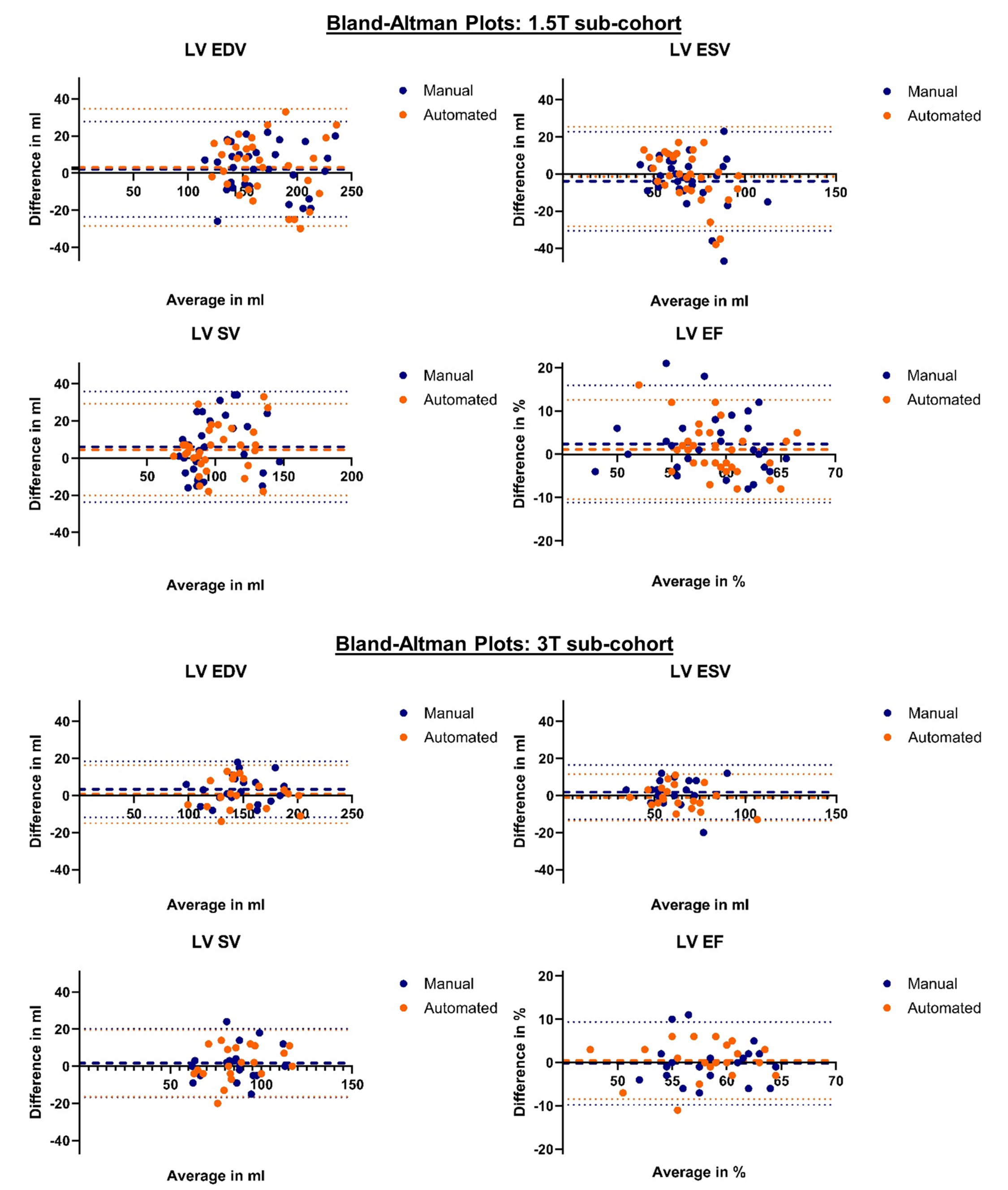
 Figure 2: Bland-Altman plots of the 1.5T and 3T sub-cohort for left ventricular volumetric parameters derived from short-axis image stacks. The matching colors thick dashed lines represent the mean bias, the matching colors dotted lines the 95% limits of agreement. LV – left ventricle; EDV – end-diastolic volume; ESV – end-systolic volume; SV – stroke volume; EF – ejection fraction.