
Rapid Fire Session

Pedro Ferreira, PhD
Senior Physicist
Royal Brompton Hospital
London, England, United Kingdom
Pedro Ferreira, PhD
Senior Physicist
Royal Brompton Hospital
London, England, United Kingdom
Sylvia Krupickova
Consultant paediatric cardiologist
Royal Brompton Hospital and Imperial College, London, United Kingdom

Julieta Rancati, MD
Paediatric Cardiologist
Royal Brompton Hospital, Hospital de Niños Pedro Elizalde & Sanatorio Franchin
Buenos Aires, Ciudad Autonoma de Buenos Aires, Argentina
Melonie Johns
Paediatric Cardiology Doctor
Royal Brompton Hospital, United Kingdom
Andrea Freixa-Benavente
Paediatric Cardiology Doctor
Royal Brompton Hospital, United Kingdom
Pekin Kaan
Paediatric Cardiology Doctor
Royal Brompton Hospital, United Kingdom

Alessandra Frigiola, MD
ACHD Consultant Cardiologist
Guy's and St Thomas' NHS Foundation Trust
London, England, United Kingdom
Hannah Bellsham-Revell, MD
Pedeatric Cardiology Consultant
Evelina London Children’s Hospital, England, United Kingdom
Kuberan Pushparajah, MD
Paediatric cardiology consultant
Evelina London Children’s Hospital/ King's College London
London, England, United Kingdom
Elettra Pomiato
Paediatric Cardiology Doctor
Royal Brompton Hospital, United Kingdom
Alberto Di Biase, MSc
Research Assistant
Imperial College London
Lonodn, England, United Kingdom
Ricardo Wage
CMR Radiographer
Royal Brompton & Harefield NHS Foundation Trust, England, United Kingdom
Camila Munoz
Research Associate
Royal Brompton Hospital and Imperial College, London, United Kingdom

Andrew D. Scott, PhD, FSCMR
Associate Professor
Imperial College London and Royal Brompton Hospital
London, England, United Kingdom
Sonia Nielles-Vallespin, PhD
Senior Lecturer
Royal Brompton Hospital and National Heart and Lung Institute, Imperial College London, United Kingdom
Dudley Pennell
Professor of cardiology
Royal Brompton Hospital and Imperial College, London, United Kingdom
 Left: Helix-angle (HA) and sheetlet-angle (E2A) distribution of values for the myocardium in the healthy LVFW (green), HLHS RVFW (blue) and LVFW (orange). Right: cDTI map examples for HA and absolute E2A in two HLHS hearts at diastole and systole.
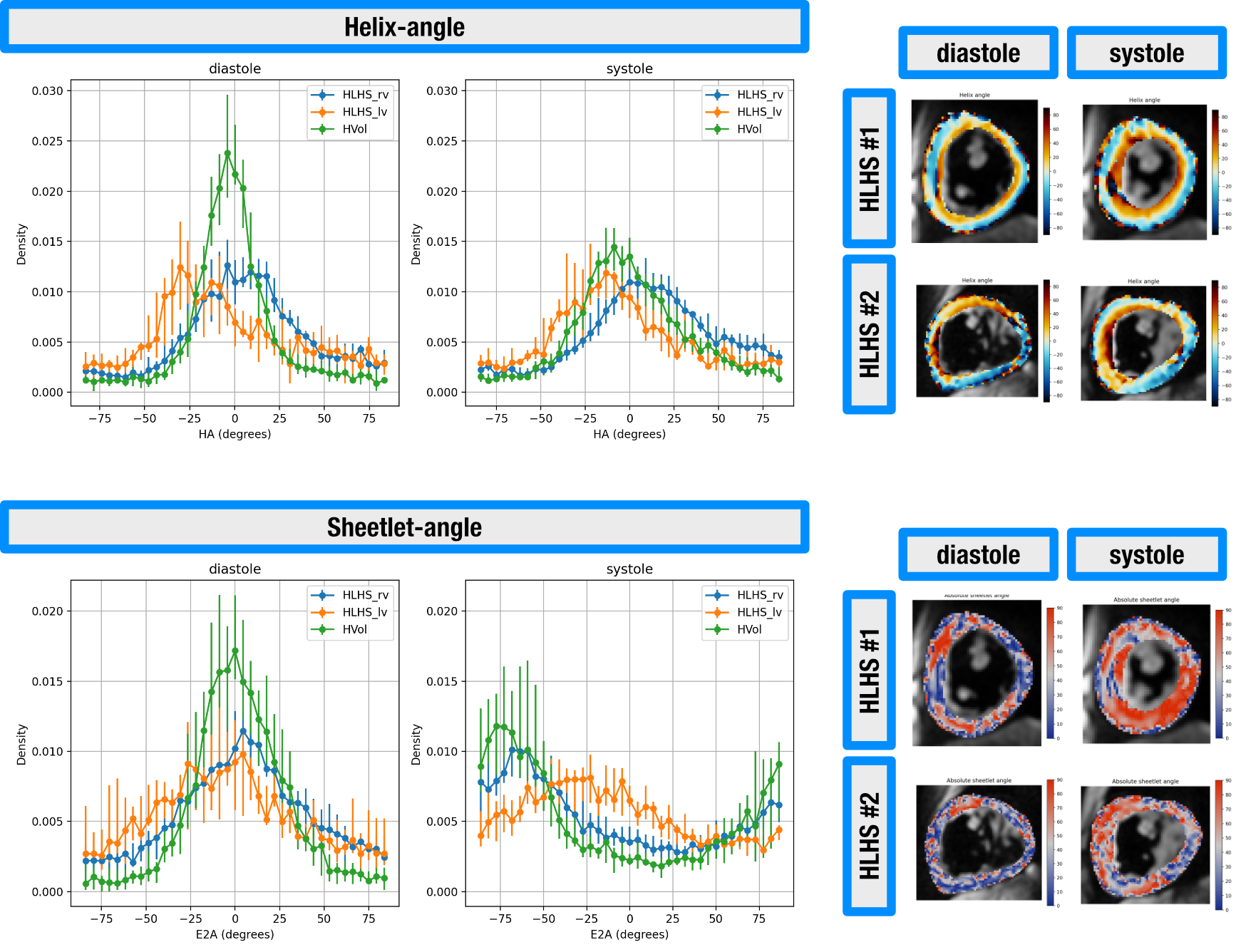 Sheetlet mobility plots: Median absolute E2A values for each subject between diastole and systole. From left to right: HLHS RVFW, HLHS LVFW, Healthy LVFW. The p-values were obtained with Mann-Whitney rank-sum and signed-rank tests.
.png)
