
Rapid Fire Session

Victor de Villedon de Naide, MSc
PhD student
Bordeaux University - INSERM U1045
Bordeaux, Aquitaine, France
Victor de Villedon de Naide, MSc
PhD student
Bordeaux University - INSERM U1045
Bordeaux, Aquitaine, France
Sane Viola
Engineer
IHU Liryc, Université de Bordeaux, France

Kalvin Narceau, MSc
Phd student
Bordeaux University - INSERM U1045
Pessac, Aquitaine, France

Thaïs Genisson, MSc
PhD student
IHU Liryc, Université de Bordeaux
Bordeaux, Aquitaine, France
Théo Richard, MSc
Engineer
IHU LIRYC, Heart rhythm disease institute, Université de Bordeaux – INSERM U1045, Avenue du Haut Lévêque, 33604, Pessac, France
Bordeaux, Aquitaine, France
Ewan Barel
Engineer
IHU Liryc, Université de Bordeaux, France

Thomas Kuestner, PhD, MSc
Professor
Medical Image and Data Analysis (MIDAS.lab), Department of Diagnostic and Interventional Radiology, University Hospital of Tübingen, 72076 Tübingen, Germany
Tübingen, Baden-Wurttemberg, Germany
Claire Bazin
MD
Centre Hospitalier Universitaire de Bordeaux, France
Edouard Gerbaud, MD, PhD
Cardiac Intensive Care Unit, Groupe Hospitalier Sud, CHU de Bordeaux, Pessac, France, France
Pierre Jaïs, MD, PhD
PROF/PhD
Hôpital Cardiologique du Haut-Lévêque, CHU de Bordeaux
Bordeaux, Aquitaine, France
Matthias Stuber, PhD
Professor/Director
CIBM/CHUV/UNIL
Lausanne, Switzerland
Hubert Cochet, MD, PhD
Professor
Bordeaux University - INSERM U1045
Bordeaux, Aquitaine, France
.jpg "Aurelien Bustin, FSCMR photo")
Aurelien Bustin, FSCMR
Research Associate
Department of Cardiovascular Imaging, Hôpital Cardiologique du Haut-Lévêque, CHU de Bordeaux, Avenue de Magellan, Pessac, France; IHU LIRYC, Electrophysiology and Heart Modeling Institute, Université de Bordeaux – INSERM U1045, Avenue du Haut Lévêque, Pessac, France; Department of Diagnostic and Interventional Radiology, Lausanne University Hospital and University of Lausanne, Lausanne, Switzerland
Bordeaux, Aquitaine, France
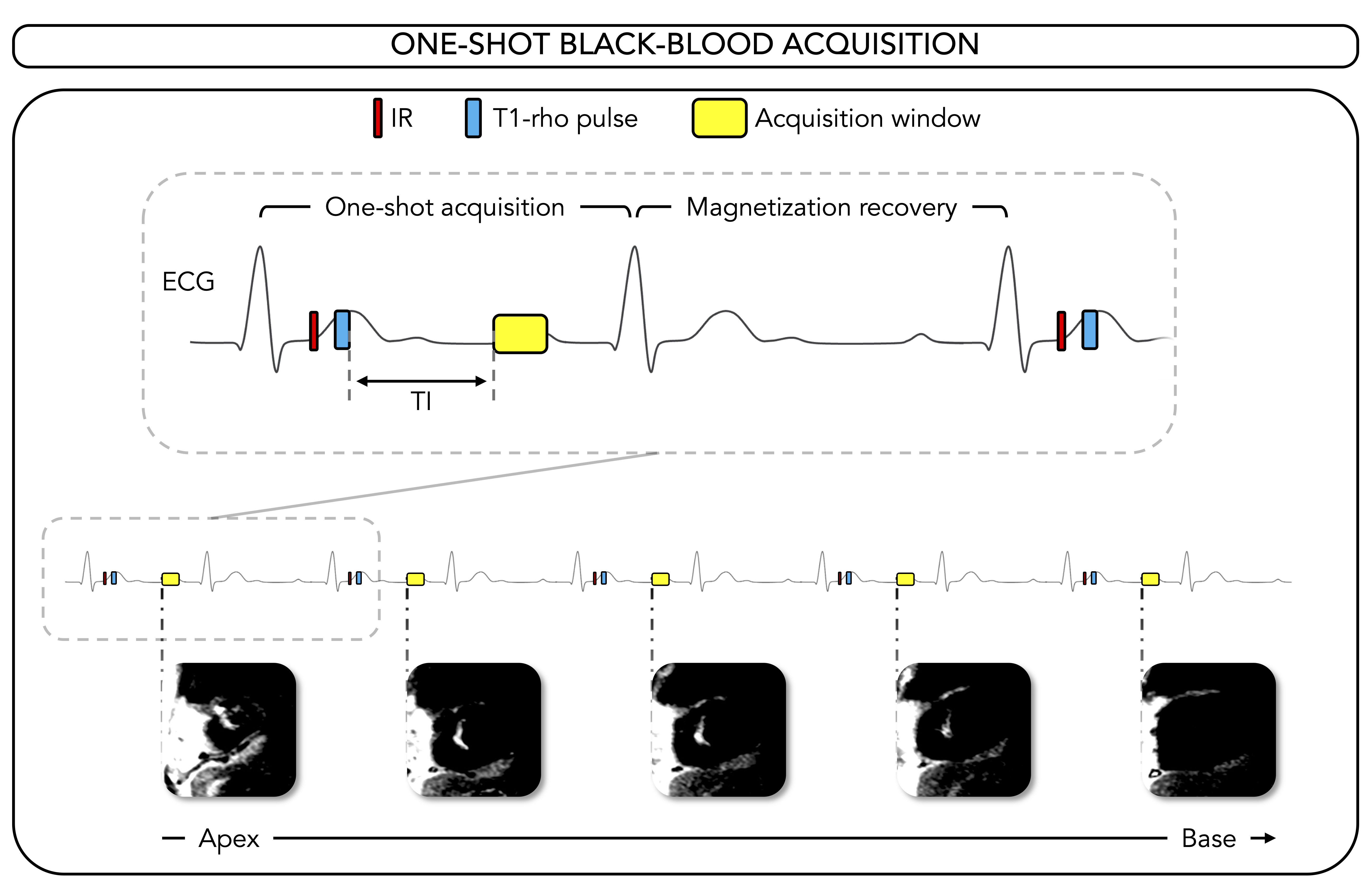 Comparison of the proposed one-shot BL with reference techniques. A) Short-axis mid-ventricular images collected with PSIR (top row), five-shots BL (second row) and the proposed one-shot BL (bottom row) in four patients with myocardial infarction. The acquisition times needed to collect each image are shown. Yellow arrows represent incertitude in LGE delineation, while red arrows represent certitude. For each patient, acquisition time was normalized to 10 short-axis slices. The shortest acquisition time was highlighted in green, otherwise in red. For one patient (Female, 62 years-old, second column, from left to right), right-ventricular infarction could be detected with BL techniques, whereas PSIR imaging, in which the hyperenhancement was confounded with blood tissue. Abbreviations: AT, acquisition time; BL, black-blood; PSIR, phase-sensitive inversion recovery.
.png) A) Comparison of scar mass assessed with five- and one-shot black-blood images through Bland-Altman analysis. B) Comparison of diagnostic confidence, following Likert scale, between PSIR, five- and one-shot black-blood images. C) Scar, remote myocardium, and blood signal intensities assessed in PSIR, five- and one-shot black-blood images. Abbreviations: PSIR, phase-sensitive inversion recovery.
A) Comparison of scar mass assessed with five- and one-shot black-blood images through Bland-Altman analysis. B) Comparison of diagnostic confidence, following Likert scale, between PSIR, five- and one-shot black-blood images. C) Scar, remote myocardium, and blood signal intensities assessed in PSIR, five- and one-shot black-blood images. Abbreviations: PSIR, phase-sensitive inversion recovery..png)
