Rapid Fire Session

Stefan K. Piechnik, PhD, FSCMR
Professor of Biomedical Imaging
University of Oxford
Oxford, England, United Kingdom
YUAN TING CHANG
Resident of Department of Radiology
Taipei Veterans General Hospital, Taiwan (Republic of China)
Chun-Ho Yun, MD
Director
Mackay Memorial Hospital
Taipei, Taipei, Taiwan (Republic of China)
Chen-Yen Chien
Attending physician
MacKay Memorial Hospital, Taiwan (Republic of China)
Wei-Ming Huang, MD
Assistant Professor
Mackay Memorial Hospital
Taipei, Taipei, Taiwan (Republic of China)
Chung-Lieh Hung
Attending physician
Mackay Memorial Hospital, Taiwan (Republic of China)

Qiang Zhang, PhD, FSCMR
Associate Professor of AI in Cardiovascular Imaging
University of Oxford
Oxford, England, United Kingdom

Ricardo A. Gonzales, PhD, FSCMR
Postdoctoral Research Fellow
Harvard Medical School
Charlestown, Massachusetts, United States
Matthew K. Burrage, MD, PhD
Research Fellow
University of Oxford, United Kingdom
.jpg "Vanessa M. Ferreira, MD, PhD, FSCMR photo")
Vanessa M. Ferreira, MD, PhD, FSCMR
Professor of Cardiovascular Medicine
University of Oxford
Oxford, England, United Kingdom
| Healthy subjects (n=25) | CAD patients (n=20) | p value |
Age (yrs) | 44.5±16.3 | 62.5±10.3 | < .0001 |
BMI (kg/m2) | 22.4±4.6 | 26.5±4.4 | 0.831 |
Male (%) | 11 (44) | 13 (65) | 0.161 |
Risk factors (%) |
|
|
|
Hypertension | 0 | 10 (50) |
|
Diabetes mellitus | 0 | 9 (45) |
|
Hypercholesterolemia | 0 | 2 (10) |
|
Family history of CAD | 0 | 3 (15) |
|
History of CAD | 0 | 11 (55) |
|
smoking | 0 | 1 (5) |
|
Medication (%) |
|
|
|
Aspirin | 0 | 13 (65) |
|
Beta-blocker | 0 | 8 (40) |
|
Statin | 0 | 14 (70) |
|
CMR clinical indices |
|
|
|
LVEF (%) | 59.2±4.4 | 62.1±7.6 | 0.147 |
LVEDVI (ml/m2) | 61.0±13.4 | 65.5±17.0 | 0.266 |
Number of remote myocardial segments& |
| 57 |
|
Number of ischemic myocardial segments# |
| 55 |
|
Number of infarcted myocardial segments# |
| 14 |
|
Invasive coronary angiography |
|
|
|
1-vessel CAD$ |
| 7 |
|
2-vessel CAD |
| 4 |
|
3-vessel CAD |
| 9 |
|
& no ischemia or infarction
# Based on results of first-pass perfusion and late gadolinium enhancement imaging
$ ≥70% stenosis in a major coronary vessel
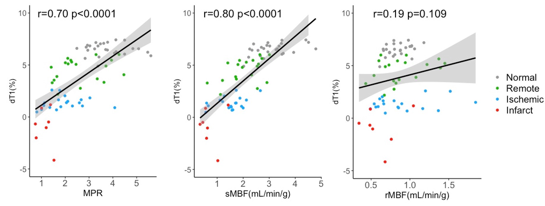
Abbreviations: CAD: coronary artery disease; CMR: cardiovascular magnetic resonance; LVEF: left ventricular ejection fraction; LVEDVI: indexed left ventricular end‐diastolic volumeMyocardial T1 values and quantitative myocardial perfusion results in healthy subjects and CAD patients
Healthy Subjects (n=25) | CAD patients (n=20) | ||||||
Normal | Remote | Ischemic | Infarct | LV blood pool | |||
Rest T1 (ms) | 934 ± 26 | 936 ± 19 | 965 ± 26 | 1146 ± 71 | 1486 ± 79 | ||
Stress T1 (ms) | 994 ± 28 | 979 ± 22 | 978 ± 27 | 1135 ± 71 | 1489 ± 83 | ||
dT1 (%) | 6.5 ± 0.6 | 4.5 ± 1.1 | 1.3 ± 0.6 | -0.9 ± 1.8 | 0.2 ± 2.8 | ||
Resting MBF | 0.9 ± 0.2 | 0.9 ± 0.3 | 0.9 ± 0.4 | 0.6 ± 0.2 | |||
Stress MBF | 3.1 ± 0.7 | 2.1 ± 0.7 | 1.5 ± 0.5 | 0.7 ± 0.4 | |||
MPR | 3.8 ± 0.8 | 2.6 ± 1.0 | 2.0 ± 0.8 | 1.1 ± 0.3 | |||
Abbreviations: CAD: coronary artery disease; dT1:delta T1; MBF: myocardial blood flow; MPR: myocardial perfusion reserveCorrelation between T1 reactivity and MPR, sMBF and rMBF. T1 reactivity is expressed as percentage change in T1 (dT1). Abbreviations: rMBF: myocardial blood flow at rest; sMBF=myocardial blood flow at stress; MPR=myocardial perfusion reserve