Rapid Fire Session

Ye Tian, PhD
Research Assistant Professor
University of Southern California
Alhambra, California, United States
Ye Tian, PhD
Research Assistant Professor
University of Southern California
Alhambra, California, United States
Jon A. Detterich, MD
Professor
Children's Hospital Los Angeles
Los Angeles, California, United States

John C. Wood, MD, PhD
Professor
University of Southern California
Los Angeles, California, United States

Krishna S. Nayak, PhD
Dean's Professor
University of Southern California
Los Angeles, California, United States
 Figure 2. Comparison of GRE, standard bSSFP, and SoS PC bSSFP in one volunteer with two 20-mm stents. End systolic phase is shown for all images. All images exhibit signal void around the stent that extends into the myocardium. GRE images suffer from poor contrast regardless of the flip angle, likely due to signal saturation of static blood, confounding the delineation of endocardial border. Standard bSSFP (phase cycle of 180o) provides improved contrast compared to GRE but with increased banding artifacts. Phase-cycled bSSFP shows a similar extent of signal void as GRE and without banding artifacts in the heart. Note that the Phase-cycled bSSFP image was acquired at a slightly different slice location than GRE and standard bSSFP, as the sequences were implemented on different console systems (Siemens and RTHawk).
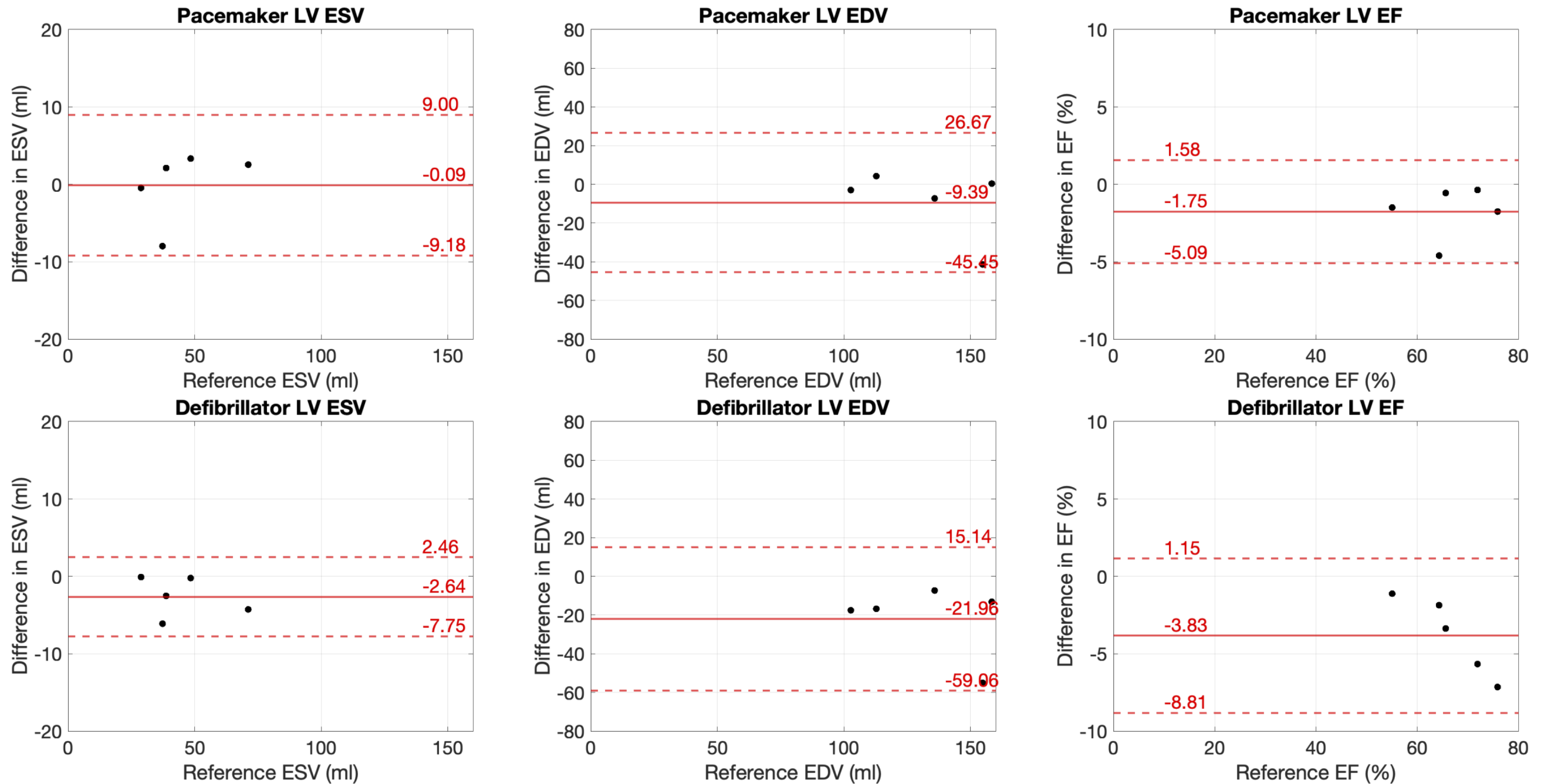
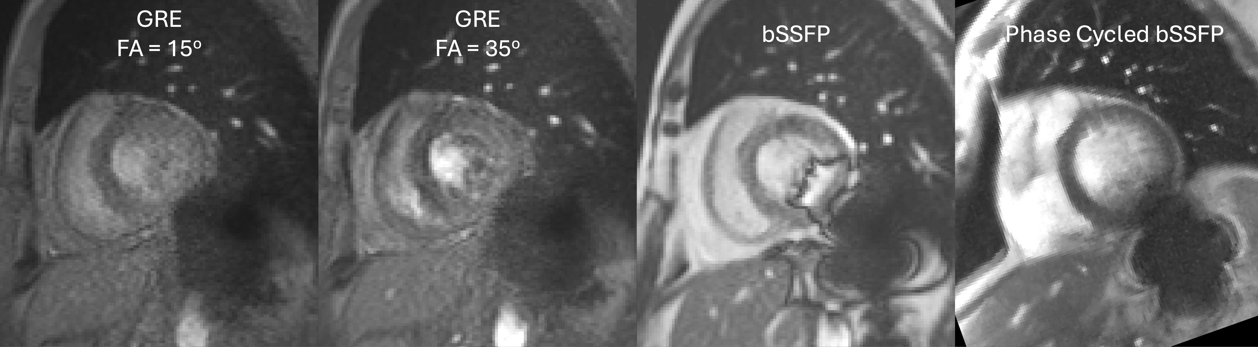 Figure 3. Comparison of left ventricular (LV) end-systolic volume (ESV), end-diastolic volume (EDV), and ejection fraction (EF) between phase-cycled spiral bSSFP (with device) and reference Cartesian bSSFP (without device). Placement of CIDs resulted in lower volume measurements compared to reference, but the differences were not statistically significant overall. For defibrillators, EDV was on average 22 mL lower than reference, likely due to image distortion. Pacemaker EF did not differ significantly from reference EF (P = 0.08), whereas defibrillator EF was significantly reduced compared to reference (P = 0.02).