Rapid Fire Session

Moses Cook, PhD
Postdoctoral Fellow
Cleveland Clinic
Cleveland, Ohio, United States
Dingheng Mai
PhD Candidate
Cleveland Clinic
Cleveland, Ohio, United States
Lipeng Ning, PhD
Assistant Professor
Brigham and Women’s Hospital, United States
Moses Cook, PhD
Postdoctoral Fellow
Cleveland Clinic
Cleveland, Ohio, United States

Zachary Player, BA, BSc
PhD Student
Case Western Reserve University, Cleveland Clinic
Cleveland Heights, Ohio, United States
Yuchi Liu, PhD
Research Scientist
Siemens Medical Solutions USA, Inc.
Cleveland, Ohio, United States

Danielle Kara, PhD
Staff Scientist
Cleveland Clinic
Cleveland, Ohio, United States
Wilson Tang, MD
Cardiologist
Cleveland Clinic, United States
Yogesh Rathi, PhD
Associate Professor of Psychiatry and Radiology
Brigham and Women’s Hospital, United States

Christopher Nguyen, PhD, FSCMR
Director, Cardiovascular Innovation Research Center
Cleveland Clinic
Cleveland, Ohio, United States
 Figure 2: (A) Traditional T1, T2, and MD maps acquired using the previously proposed sequence and acquisition protocol.4 Spatially averaged histograms of these parameters are shown for two regions of interest—scar and remote—as labeled on the T1 map. Clear shifts in the distributions were observed between scar and remote tissue, with the scar region showing elevated T1, T2, and MD values. (B) Spatially averaged DRSI results in the same two regions. In the remote (non-infarcted) region, two distinct well-resolved peaks were identified, and the dominant peak have D, R1, and R2 values comparable to those in the traditional maps. In contrast, the enhanced (scar) region exhibited multiple peaks in the DRSI spectra, with the dominant component showing reduced R1 and R2 and elevated D, consistent with MI pathology. Similar characteristics to remote tissue were observed, though it’s hard to distinguish from the conventional maps.
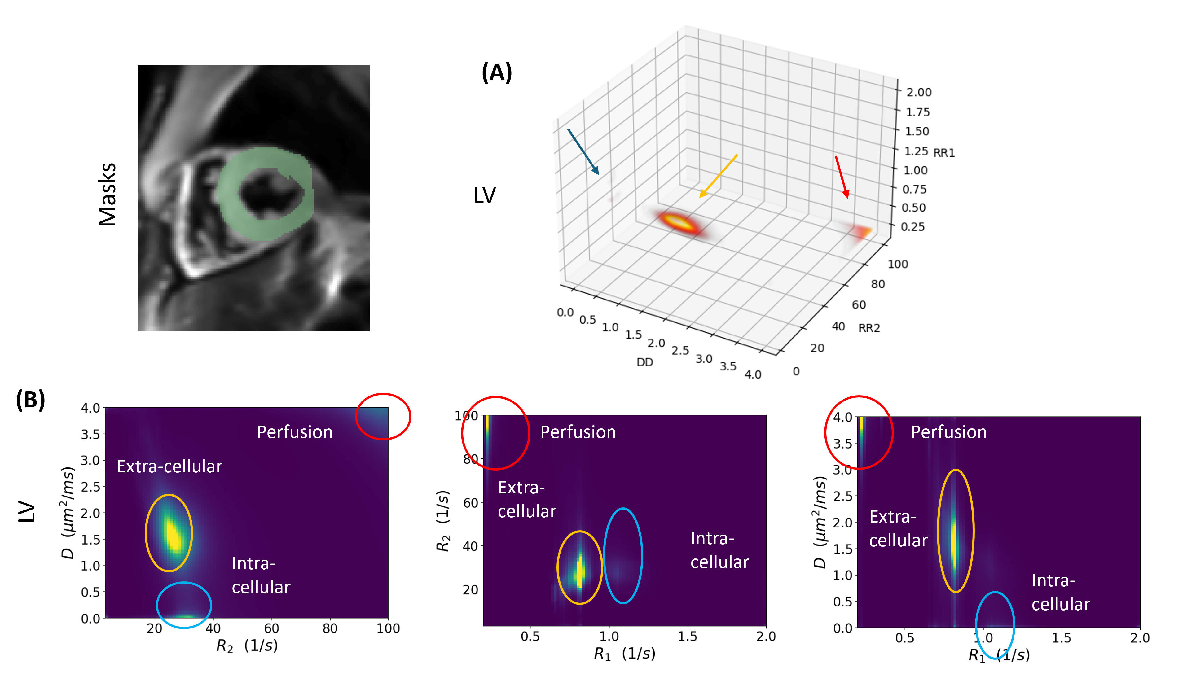
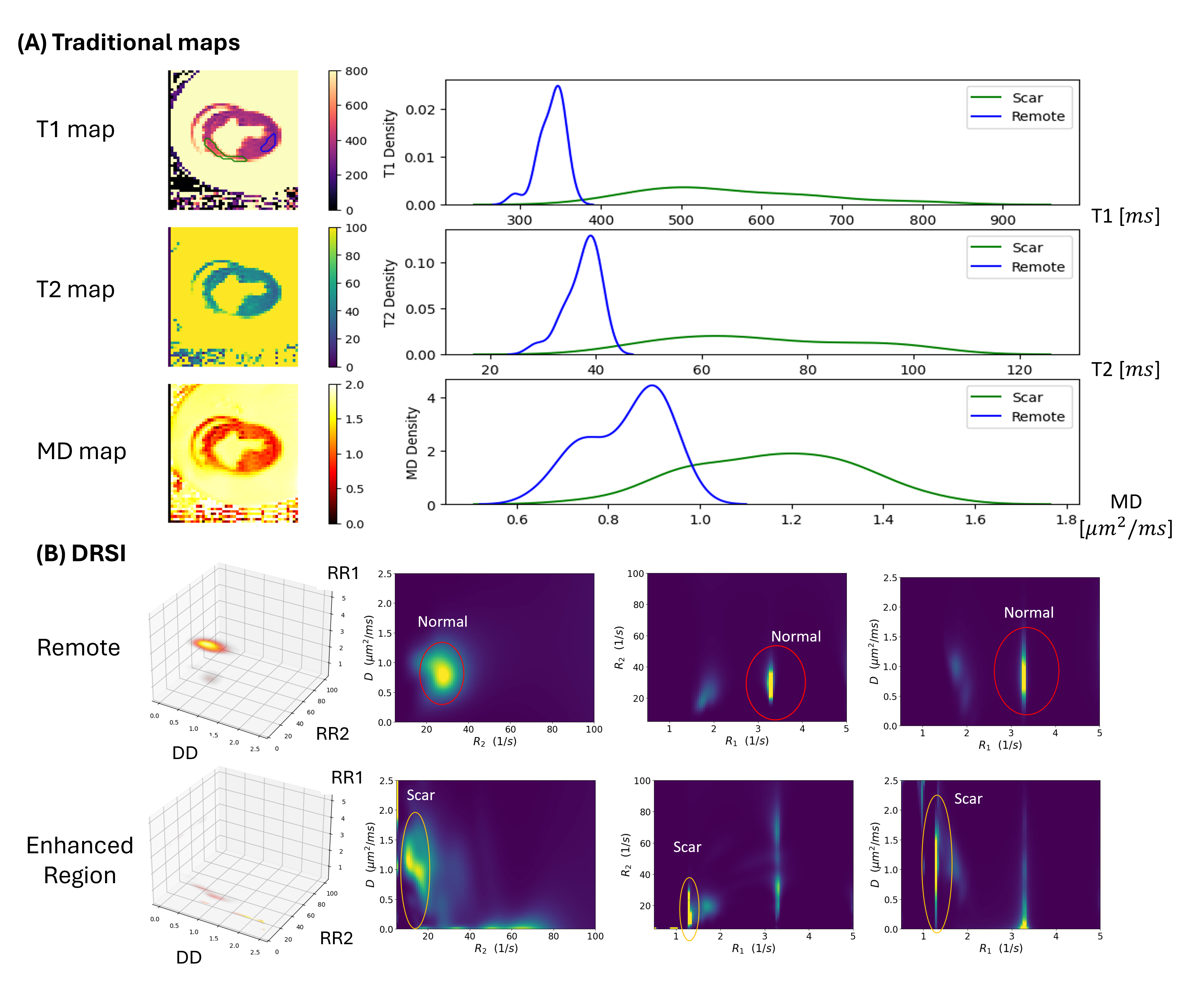 Figure 3: Spatially averaged 3D diffusion-relaxation (D–R1–R2) distributions estimated using the MaxEnt method for the left ventricle (LV) from a representative mid-ventricular slice for a healthy volunteer. Three distinct peaks were observed: extra-cellular (orange, moderate D, R1, and R2), intra-cellular (blue, zero D), and perfusion-related (red, very high D). (A) 3D plots of the distributions in D–R1–R2 space, with arrows indicating three primary tissue components. (B) 2D projections onto the D–R2, R1–R2, and D–R1 planes. The extra-cellular component resembles those observed in the remote zone of the ex-vivo heart and shows values comparable to T1, T2, and MD values in the healthy normal range. Elongation of extra-cellular component along the D-axis in the D–R1 projection suggests that the current diffusion sampling scheme is sparse and requires further optimization. The intra-cellular component observed in-vivo might be missing in the ex-vivo setting due to ultra-high R1, and perfusion component is unique for in-vivo scan only.