Early Career Award Session
Virtual Recording

Kalvin Narceau, MSc
Phd student
Bordeaux University - INSERM U1045
Pessac, Aquitaine, France
Kalvin Narceau, MSc
Phd student
Bordeaux University - INSERM U1045
Pessac, Aquitaine, France

Thaïs Genisson, MSc
PhD student
IHU Liryc, Université de Bordeaux
Bordeaux, Aquitaine, France

Victor de Villedon de Naide, MSc
PhD student
Bordeaux University - INSERM U1045
Bordeaux, Aquitaine, France
Théo Richard, MSc
Engineer
IHU LIRYC, Heart rhythm disease institute, Université de Bordeaux – INSERM U1045, Avenue du Haut Lévêque, 33604, Pessac, France
Bordeaux, Aquitaine, France

Alexis Jacquier, MD, PhD
Prof
Aix Marseille Univ, CNRS, CRMBM, Marseille, France; APHM, Hôpital Universitaire Timone, Radiology Dept, Marseille, France
Marseille, Provence-Alpes-Cote d'Azur, France
Axel Bartoli, MD
Medical doctor
Aix-Marseille Université, France
Salim Si-Mohamed, MD
Medical Doctor
Louis Pradel Hospital, Hospices Civils de Lyon; University of Lyon, France
Matthias Stuber, PhD
Professor/Director
CIBM/CHUV/UNIL
Lausanne, Switzerland
Hubert Cochet, MD, PhD
Professor
Bordeaux University - INSERM U1045
Bordeaux, Aquitaine, France
.jpg "Aurelien Bustin, FSCMR photo")
Aurelien Bustin, FSCMR
Research Associate
Department of Cardiovascular Imaging, Hôpital Cardiologique du Haut-Lévêque, CHU de Bordeaux, Avenue de Magellan, Pessac, France; IHU LIRYC, Electrophysiology and Heart Modeling Institute, Université de Bordeaux – INSERM U1045, Avenue du Haut Lévêque, Pessac, France; Department of Diagnostic and Interventional Radiology, Lausanne University Hospital and University of Lausanne, Lausanne, Switzerland
Bordeaux, Aquitaine, France
 Figure 2: Visual comparison of LGE, T1rho (500Hz), T1rho (0Hz), and MDI maps in 9 patients. For each patient, three short-axis slices (1.5x1.5x8 mm) were acquired. The MDI maps derived from the proposed sequence show clear regional elevations corresponding to LGE-positive regions, despite visible noise influence in some cases.
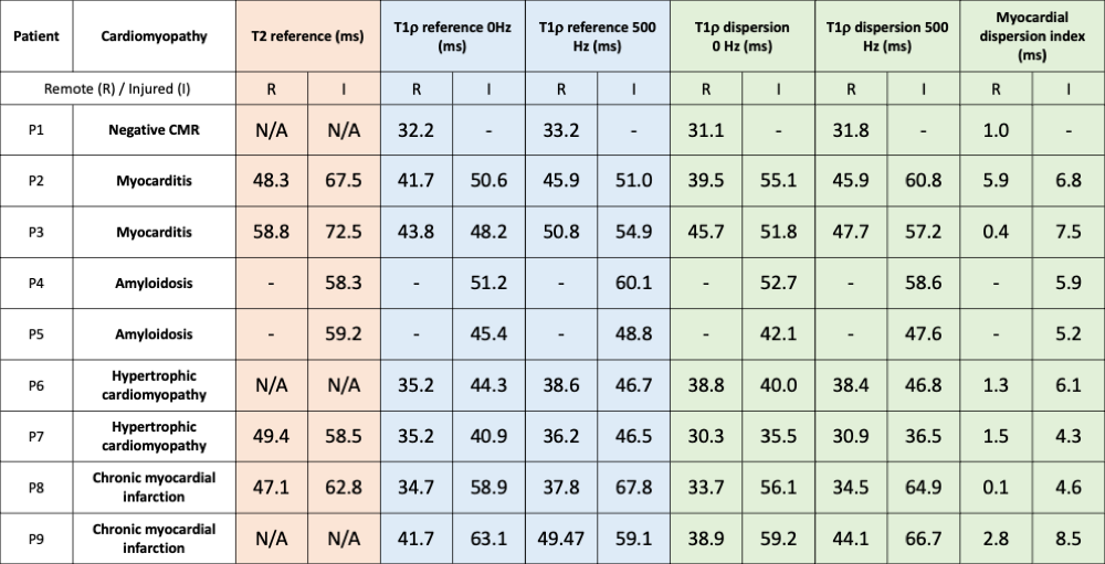
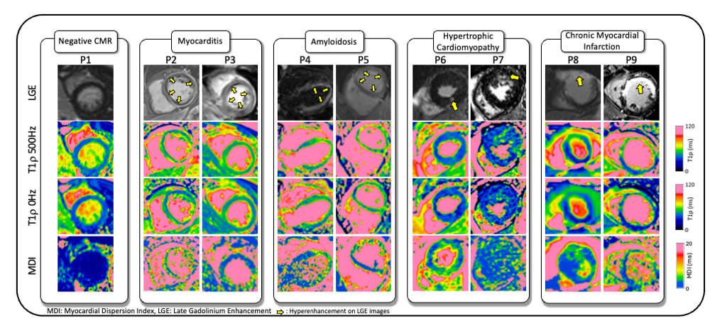 Figure 3: ROI-based comparison of remote and injured myocardium across mapping techniques. T2 maps (when available), T1rho (500Hz and 0Hz) maps, MDI maps from the proposed sequence, and reference T1rho maps were analyzed. With the exception of P2 (diffuse myocarditis, unusually high remote values) and diffuse amyloidosis cases (no true remote myocardium), remote myocardium values clustered around 1.0-2.8 ms, while injured myocardium ranged from 4.3-8.5 ms.