Rapid Fire Session

Raymond Kim, MD
Professor
Duke Cardiovascular Magnetic Resonance Center
Durham, North Carolina, United States
Céleste Chevalier, MD
Postdoctoral fellow
Duke University Medical Center
Durham, North Carolina, United States
Michele Parker, MSc
Statistician / Business Manager
Duke Cardiovascular Magnetic Resonance Center, North Carolina, United States

David C. Wendell, PhD
Senior Research Scientist
Duke University Medical Center
Durham, North Carolina, United States
Han Kim, MD
Associate Professor of Medicine
Duke University Medical Center
Durham, North Carolina, United States
Chen Enn-Ling, PhD
Assistant Professor in Medicine
Duke University Medical Center, United States
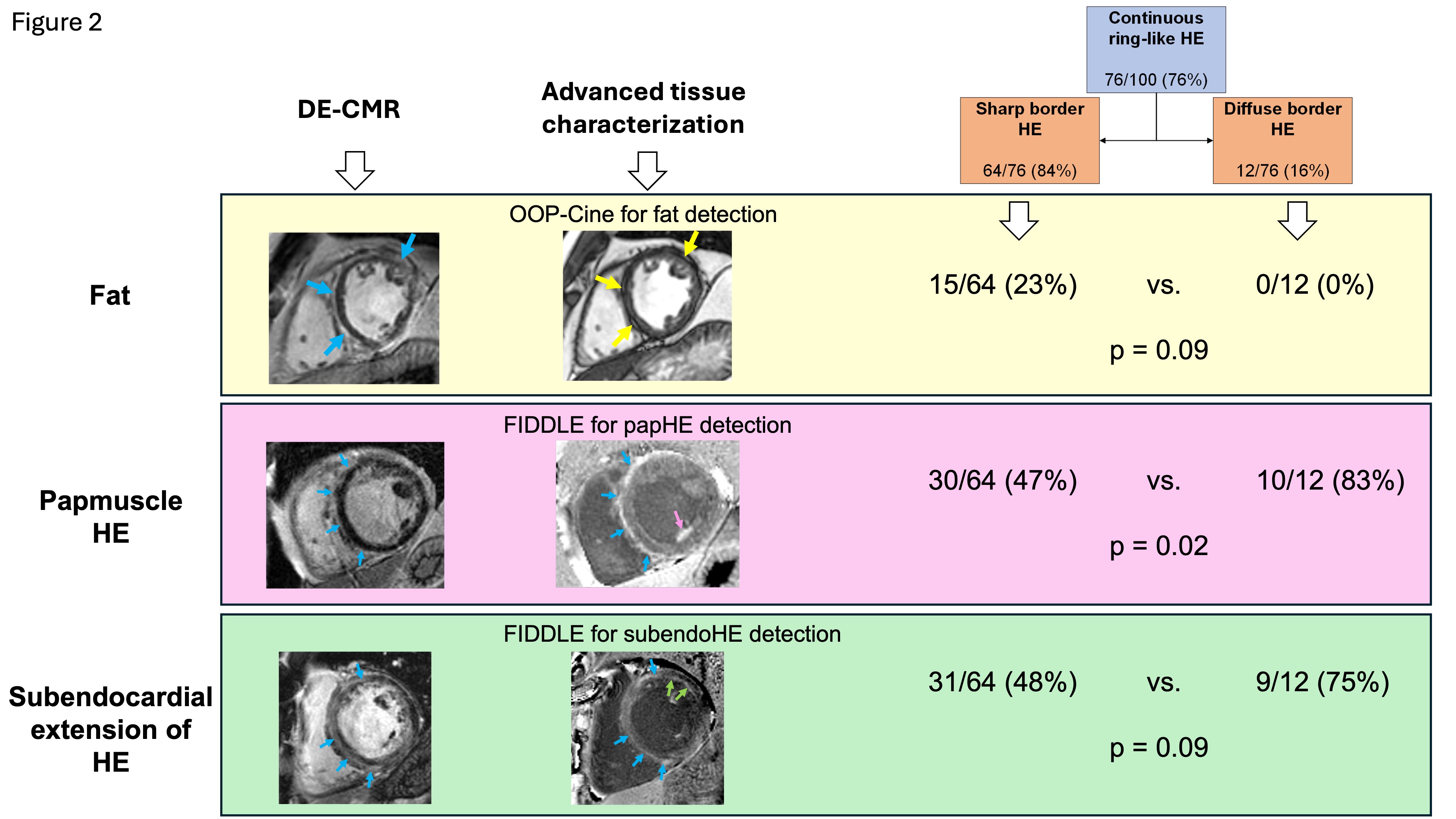
 Figure 2. Advanced CMR tissue characterization in patients with continuous ring-like hyperenhancement (HE). Yellow box: Delayed-enhancement (DE) images show a sharp border striae of HE in the septum and anterolateral wall (blue arrows). The patient‘s corresponding out-of-phase (OOP) cine images, which are sensitive to detecting fatty metaplasia, demonstrate that the HE is predominantly secondary to intramyocardial fat (India ink appearance, yellow arrows). Overall, only patients with sharp border striae had associated fatty metaplasia. Pink box: Flow-independent dark-blood delayed enhancement (FIDDLE) imaging (right) demonstrates papillary muscle scarring (pink arrow) as well as an epicardial ring of HE which also involves the RV side of the septum (blue arrows). Green box: FIDDLE image shows a midwall ring of HE in the septum (blue arrows) with continuation of the ring to the subendocardial layer of the anterolateral wall (green arrows). Those with diffuse border rings were moderately more likely to have subendocardial extension of the ring and papillary muscle scarring.