Congenital Track
Virtual Recording

Amol A. Kulkarni, MD
Junior Consultant
Jupiter Hospital, Thane
Thane, Maharashtra, India
Amol A. Kulkarni, MD
Junior Consultant
Jupiter Hospital, Thane
Thane, Maharashtra, India
Priya D. Chudgar, MD, FSCMR
Cardiac Radiologist
Jupiter Hospital
Mumbai, Maharashtra, India
Nitin Burkule, MD, FSCMR
Cardiologist
Jupiter Hospital
Thane, Maharashtra, India
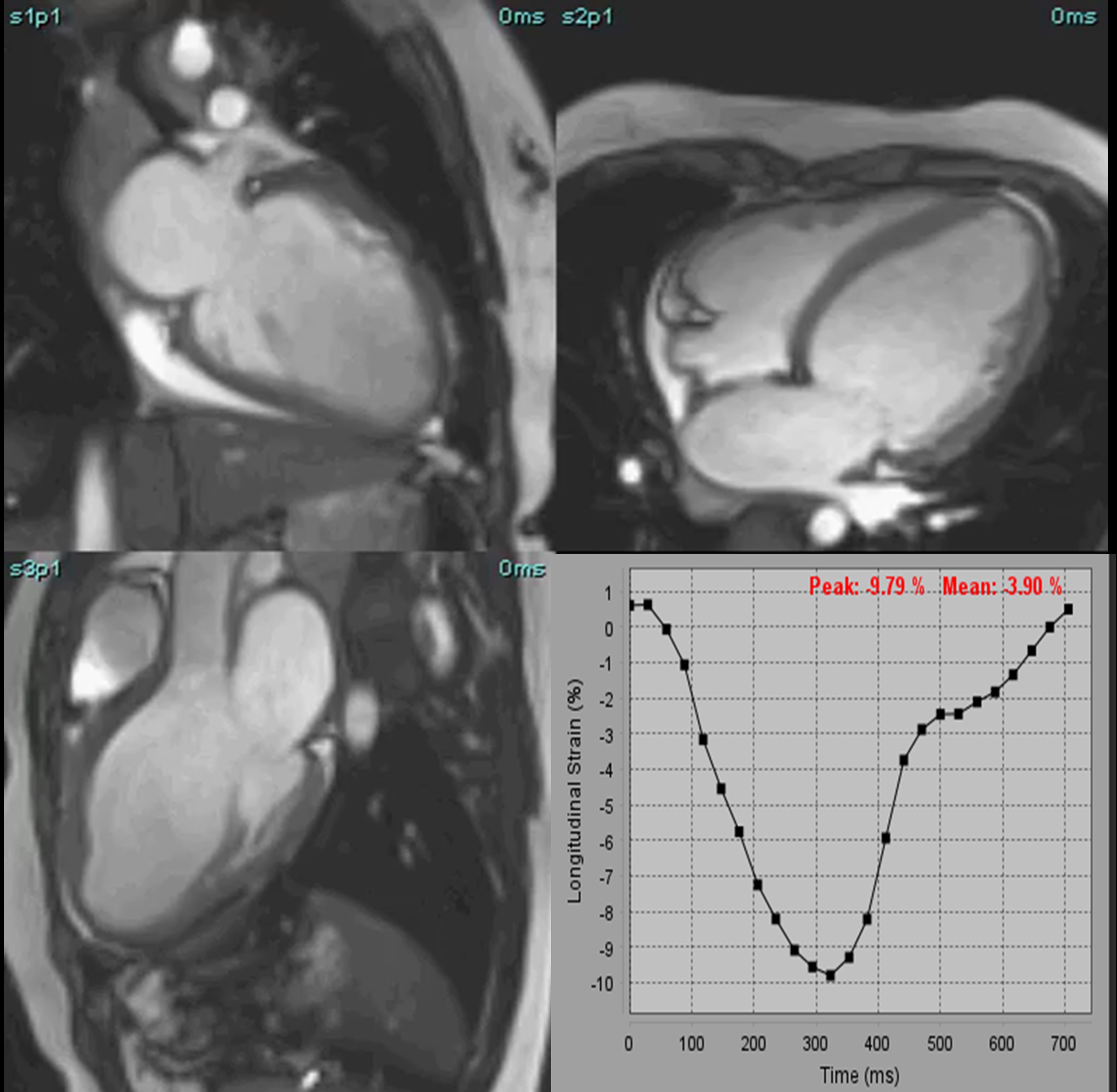 (A) Precontrast and (B) Post contrast T1 mapping shows elevated global native T1 values 1387 ms. (C) T2 mapping shows regional elevation of T2 values at the mid anterior wall ~ 52 ms, Short axis PSIR images at show subendocardial to focal transmural LGE at base (D), subepicardial LGE at mid (E) and apical cavity (F) - blue arrowheads.
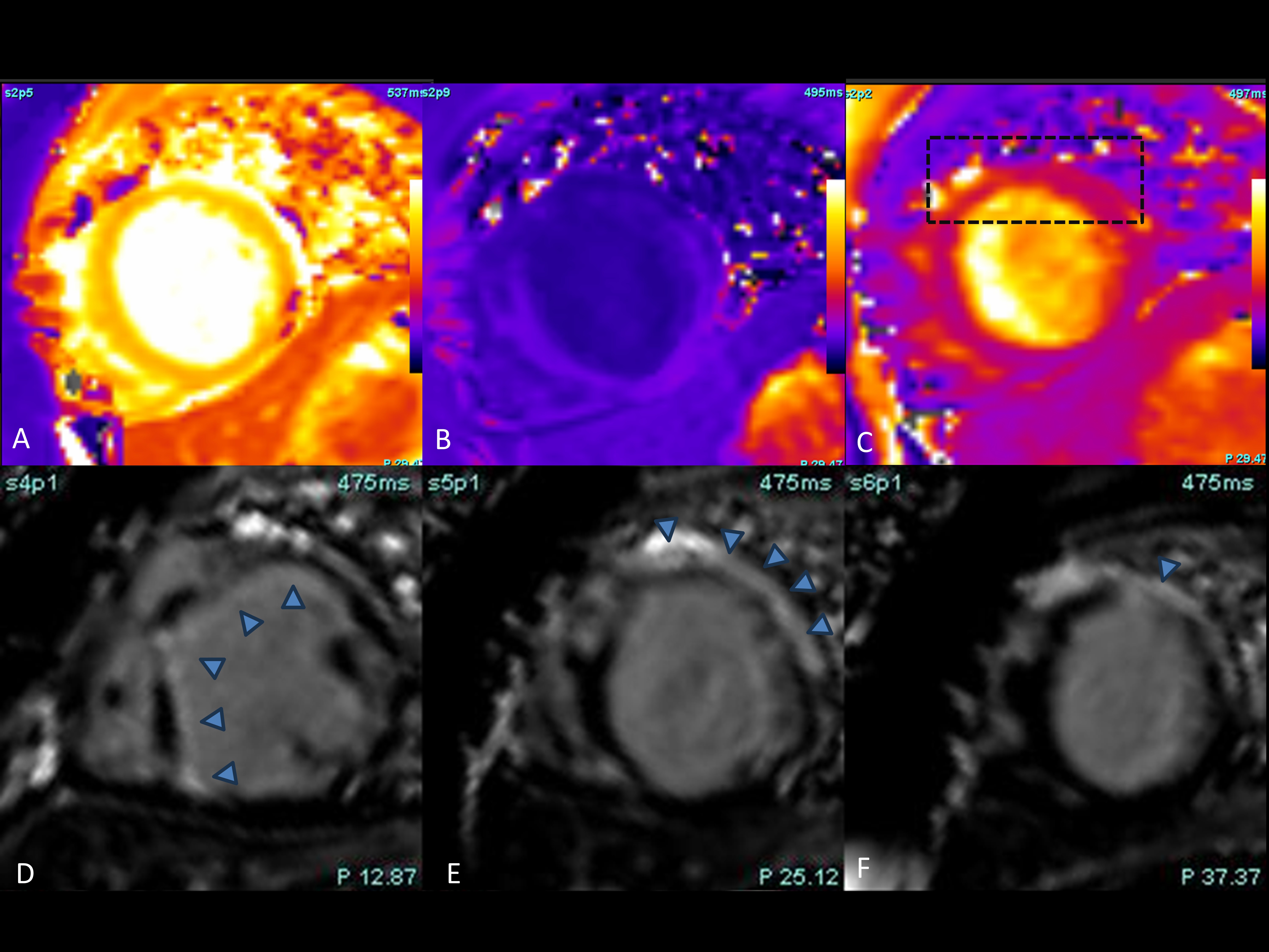 Axial PET-CT images show FDG avid enlarged lymph nodes (Blue arrows) at right axillary, right highest mediastinal and subcarinal nodal stations.
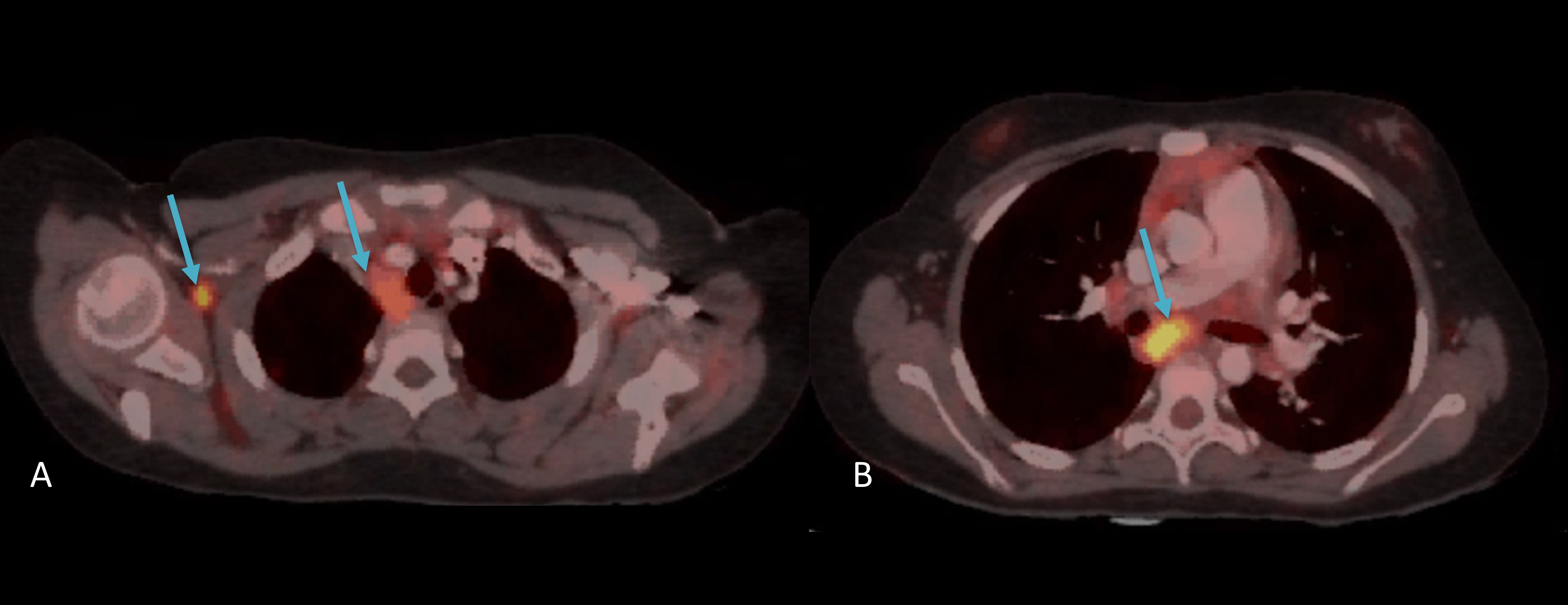