Congenital Track
Virtual Recording
Takegawa Yoshida, MD
Associate Project Scientist
University of California, Los Angeles
Los Angeles, California, United States
J. Paul Finn, MD, MSc
Professor
University of California, Los Angeles
Los Angeles, California, United States

Shu-Fu Shih, PhD
Assistant Project Scientist
University of California, Los Angeles
Los Angeles, California, United States

Kim-Lien Nguyen, MD
Associate Professor of Cardiovascular Medicine and Radiology
David Geffen School of Medicine at UCLA and VA Greater Los Angeles Healthcare System
Los Angeles, California, United States
Arash Bedayat, MD
Assistant Professor
University of California, Los Angeles
Los Angeles, California, United States
Takegawa Yoshida, MD
Associate Project Scientist
University of California, Los Angeles
Los Angeles, California, United States
Takegawa Yoshida, MD
Associate Project Scientist
University of California, Los Angeles
Los Angeles, California, United States
Ning Jin, PhD
Senior Key Expert
Siemens Healthineers
Cleveland, Ohio, United States

Xiaoming Bi, PhD
Director, Cardiovascular MR Collaborations
Siemens Medical Solutions USA, Inc.
Oak Park, California, United States
Fei Han
Siemens Healthineers
Siemens Healthineers, United States

Xiaodong Zhong, PhD
Associate Professor
University of California, Los Angeles
Los Angeles, California, United States
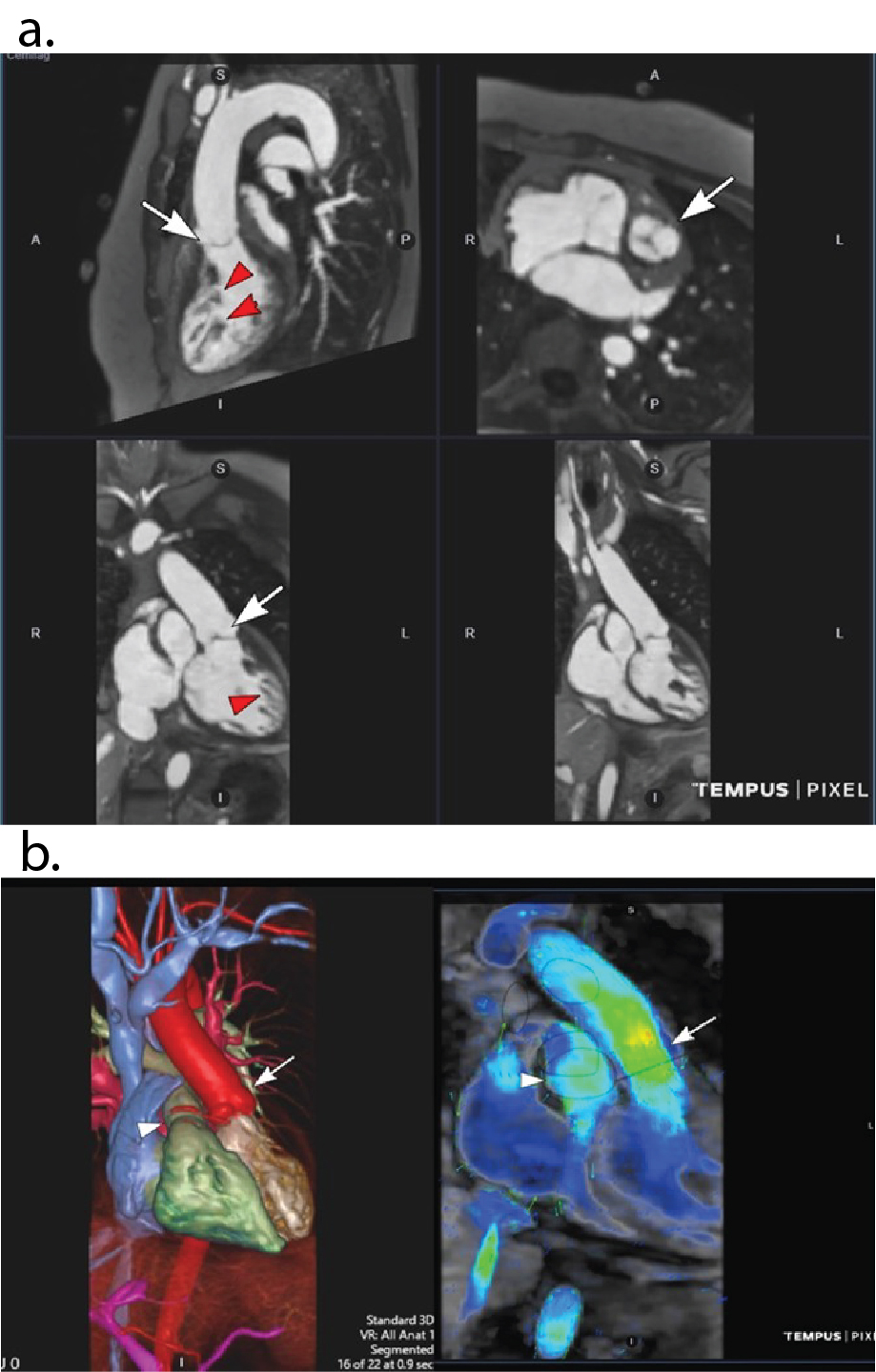 Figure 2: 42 y.o. female with mild aortic coarctation. a) Multiplanar reconstructions on OsiriX software displays one of 10 phases from BH 4D MUSIC at 3.0T . Breath-hold duration 22 secs with acceleration factor X15. Note the uniform blood signal and clear definition of the coarctation (white arrow) and left coronary artery (red arrow). b) Same patient and same acquisition as in Fig 4a. Color volume rendered frame from BH 4D MUSIC (left panel; Vitrea workstation) and corresponding 4D flow (right panel; Tempus cloud) on one of 10 phases shows the anatomy and flow field of the coarctation (arrow). c) Same patient as in Figs 2a,b. Separate BH 4D MUSIC acquisition with higher temporal resolution for full coverage cine imaging. Breath-hold duration 19 secs with acceleration factor X15. Top panel shows single phase from 2D cine at several short axis positions. Bottom panel shows corresponding short axis cine 18 phase reconstructions from BH 4D MUSIC with 3mm contiguous slices and 50 ms temporal resolution.
 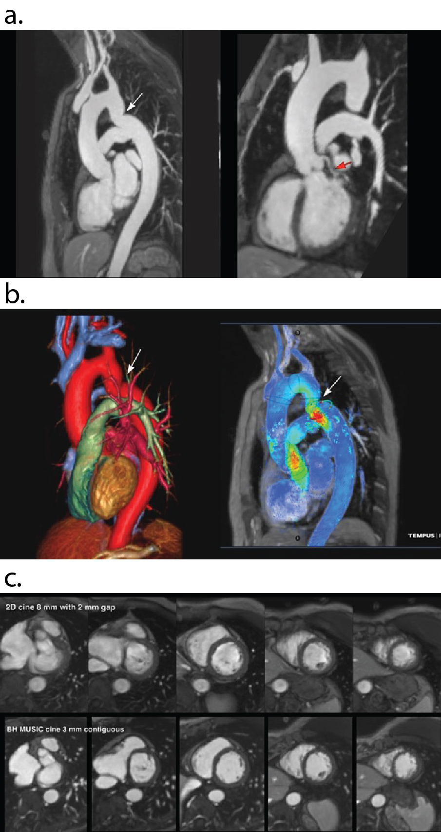 Figure 3: a) Bland-Altman plots comparing left and right ventricular end systolic and end diastolic volumes on BH 4D MUSIC and BH 2D cine . Mean values were highly similar between 4D MUSIC and 2D cine, with a mean difference less than 2 ml and a spread less than 6 ml. b) Linear regression plots of left ventricular end diastolic volume (LVEDV) and end systolic volumes (LVESV) and corresponding right ventricular volumes (RVEDV, RVESV) on BH 4D MUSIC (y axis) and BH 2D cine (x axis) in 30 adult patients with congenital heart disease. The correlation is very high, with most values lying close to the line of identity.
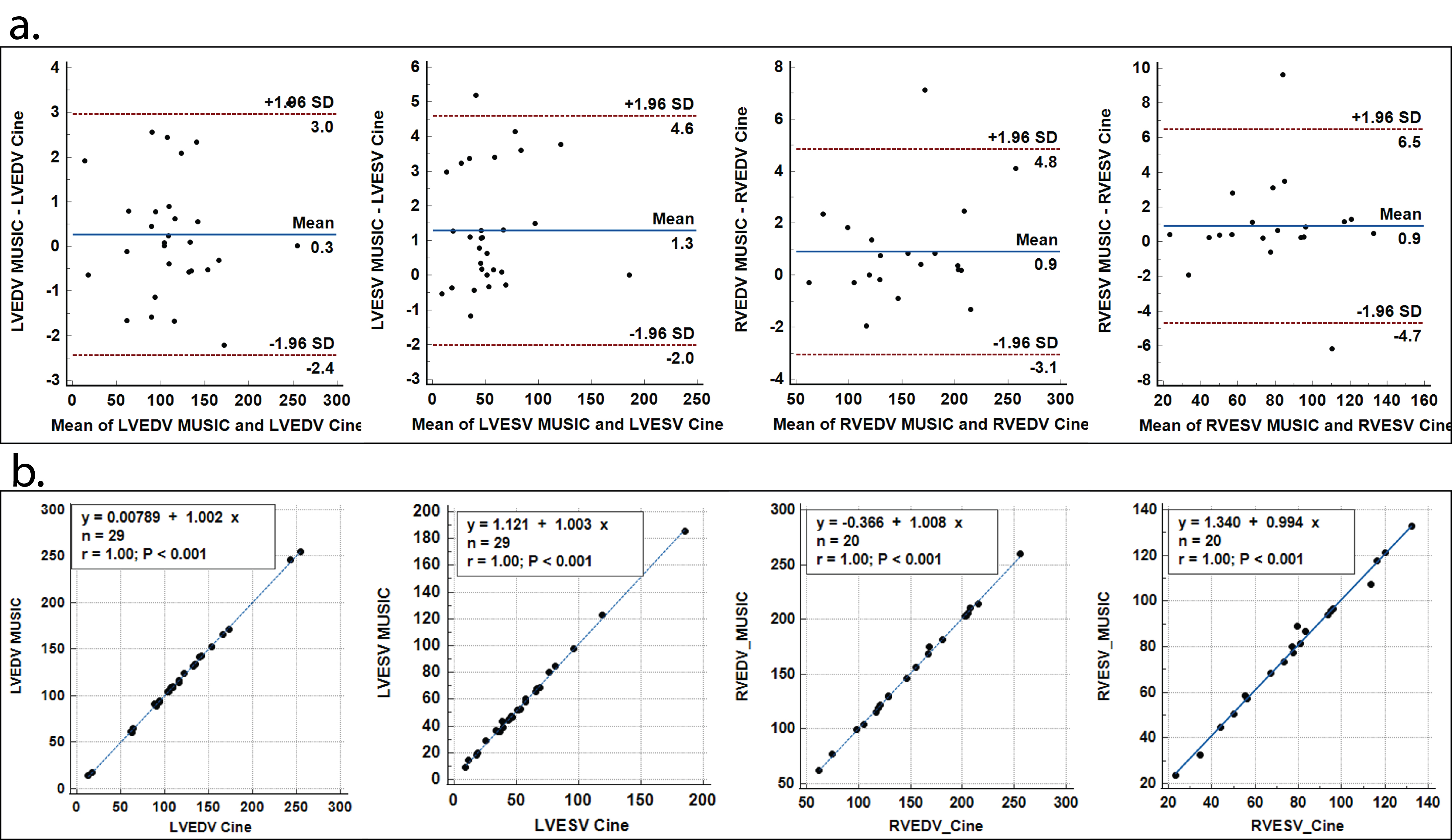