Quick Fire Session

Miguel Angel Valdes-Camaño, MD
Internal Medicine, Cardiology, Echocardiography and Cardiovascular Imaging
Hospital Chiriqui
David, Chiriqui, Panama
Miguel Angel Valdes-Camaño, MD
Internal Medicine, Cardiology, Echocardiography and Cardiovascular Imaging
Hospital Chiriqui
David, Chiriqui, Panama
 Figure 2. a: Initial transthoracic echocardiogram (TTE), left ventricle (LV) apical 5-chamber view, P2 prolapse (arrow). b: TTE, LV apical 4-chamber view, color Doppler mode, mitral regurgitation (MR) flows (asterisks). c and d: cardiac magnetic resonance (CMR), cine sequences in 3-chamber views in diastole (c) and systole (d); in systole, the anterior mitral leaflet SAM (arrow) and MR flow (asterisks) can be seen. e: CMR, inversion recovery sequence, 4-chamber view with anteroseptal and apical transmural late gadolinium enhancement (asterisks).
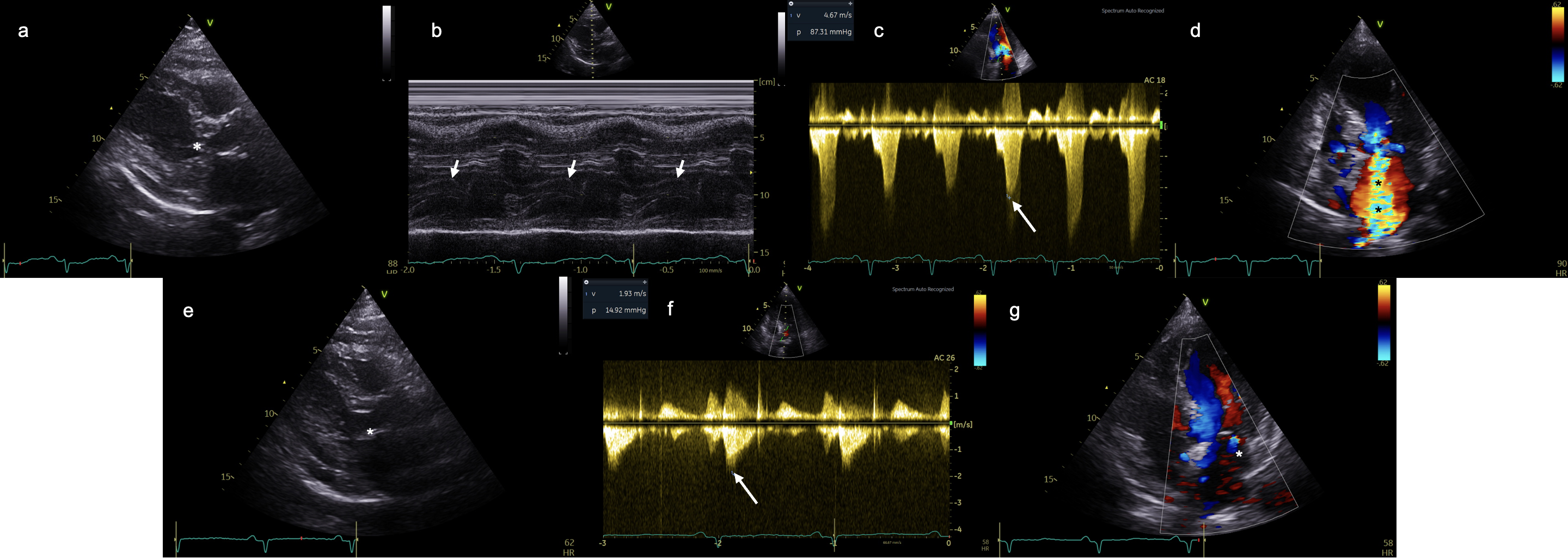
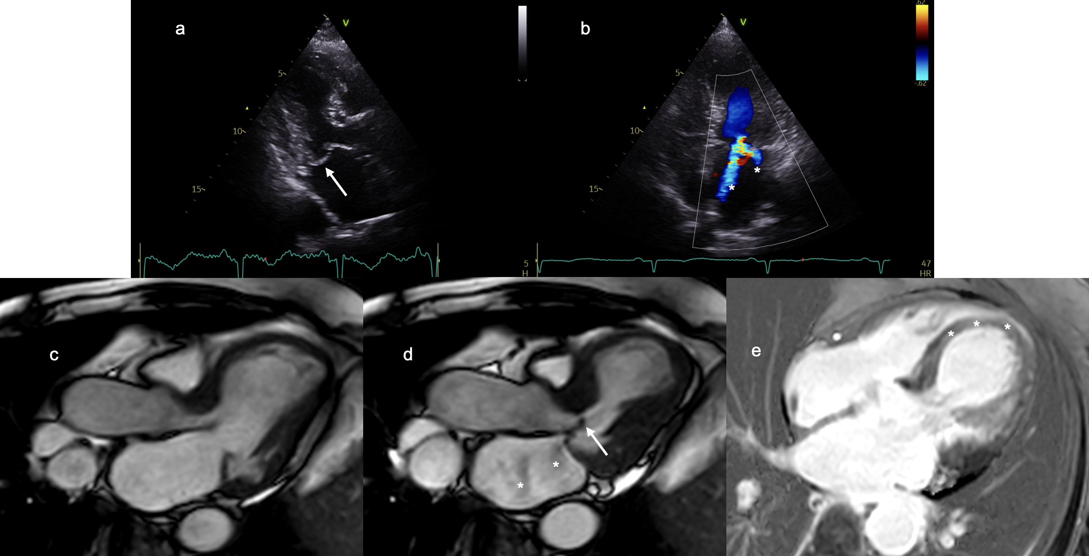 Figure 3. a, b, c, and d: Second TTE. a: LV parasternal long-axis view (PLAX) with SAM of the anterior mitral leaflet (asterisk). b: M-mode LV PLAX, SAM of the anterior mitral leaflet (arrows). c: LV apical 5-chamber view, continuous-wave Doppler, obstructive LV outflow tract (LVOT) gradient with scimitar morphology and maximum gradient of 87.3 mmHg (arrow). d: LV apical 4-chamber view, color Doppler, increasing MR severity to severe compared with the initial TTE (asterisks). e, f, and g: Predischarge TTE. e: LV PLAX, without evidence of SAM (asterisk). f: LV apical 5-chamber view, continuous-wave Doppler, with resolution of the obstructive gradient (arrow). g: LV apical 4-chamber view, color Doppler mode, with decreased severity of MR to mild (asterisk).