Quick Fire Session

Javier De La Cruz Pelayo, MD
Cardiovascular Imaging Fellow
National Institute of Cardiology
Mexico City, Distrito Federal, Mexico
Javier De La Cruz Pelayo, MD
Cardiovascular Imaging Fellow
National Institute of Cardiology
Mexico City, Distrito Federal, Mexico
Miguel Cruz, MD
Medical physician of cardiac magnetic resonance
National Institute of Cardiology "Ignacio Chávez"
CDMX, Distrito Federal, Mexico
Aloha Meave gonzalez, MD
Chair cardiac magnetic resonance
Instituto Nacional de Cardiología Ignacio Chávez
Mexico city, Distrito Federal, Mexico
José Martín Alanís Naranjo, MD
Cardiovascular Imaging Fellow
Instituto Nacional de Cardiología Ignacio Chávez
Mexico City, Mexico City, Mexico
María Mónica De Ávila Gómez, MD
Cardiovascular Imaging Fellow
National Institute of Cardiology
Ciudad de Mexico, Distrito Federal, Mexico
Gennaro Basso Barba, MD
Cardiovascular Imaging Fellow
National Institute of Cardiology, Distrito Federal, Mexico
Daniel Cordova Galván
Resident physician in Diagnostic Radiology
National Institute of Cardiology, Distrito Federal, Mexico
 Fig. 2 4D Flow in the pulmonary artery: A) Pathline visualization demostrating flow acceleration along the anterior wall of the main pulmonary artery and turbulent flow at the origin of the left pulmonary artery (LPA). B) Regions of increased shear wall stress highlighted (red arrows). C) Quantitive analysis in pascals (Pa) comparing both pulmonary branches. D-F) Multiplanar reconstructions depicting the site of device protrusion into the proximal LPA.
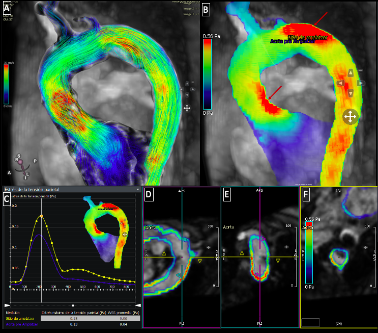
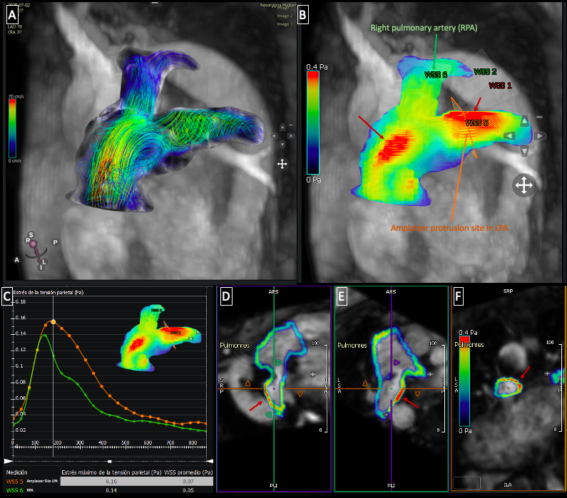 Fig. 3 4D Flow in the aorta: A) Pathline visualization demostrating flow acceleration in the ascending aorta and turbulent flow at the site of device protusion. B) Regions of increased wall shear stress (red arrows). C) Quantification in pascals (Pa) comparing wall shear stress before the device and at the device site. D-F) Multiplanar reconstructions illustrating the location and extent of the device protrusion within the aortic lumen.