Quick Fire Session
LUCIA RODRIGUEZ EYRAS, MD
Cardiologist. Advanced cardiac imaging
Hospital Universitari Vall d'Hebron
Mar del Plata, Buenos Aires, Argentina
LUCIA RODRIGUEZ EYRAS, MD
Cardiologist. Advanced cardiac imaging
Hospital Universitari Vall d'Hebron
Mar del Plata, Buenos Aires, Argentina
Luciano Martin Brown, MD
Cardiologist
Hospital Dr. Oscar Alende, Clinica Colon Mar del Plata, Argentina
Ana L. Tufare, PhD
Cardiologist and Echocardiography Specialist, Head of Congenital Heart Disease Department
HIGA Dr. O. Alende, Mar del Plata
Mar del Plata, Buenos Aires, Argentina
| Parameter | Value |
|---|---|
| Patient | 23 y, 80 kg, 185 cm, BSA 2.03 m² |
| Ventricle | Single ventricle with vestigial RV |
| SV EDV / ESV | 220 / 100 ml (108 / 48 ml/m²) |
| SV EF | 55% |
| LA / RA | 34 ml / 7 cm² |
| Septum thickness | 20 mm |
| RV vestigial free wall thickness | 18 mm |
| Mitral valve | Elongated anterior leaflet |
| T1 native septal | 1027 ms |
| Late gadolinium enhancement | Mid–apical septum & vestigial RV wall, heterogeneous |
| Fontan conduit | Extracardiac, permeable |
| Situs | Solitus, mesocardia |
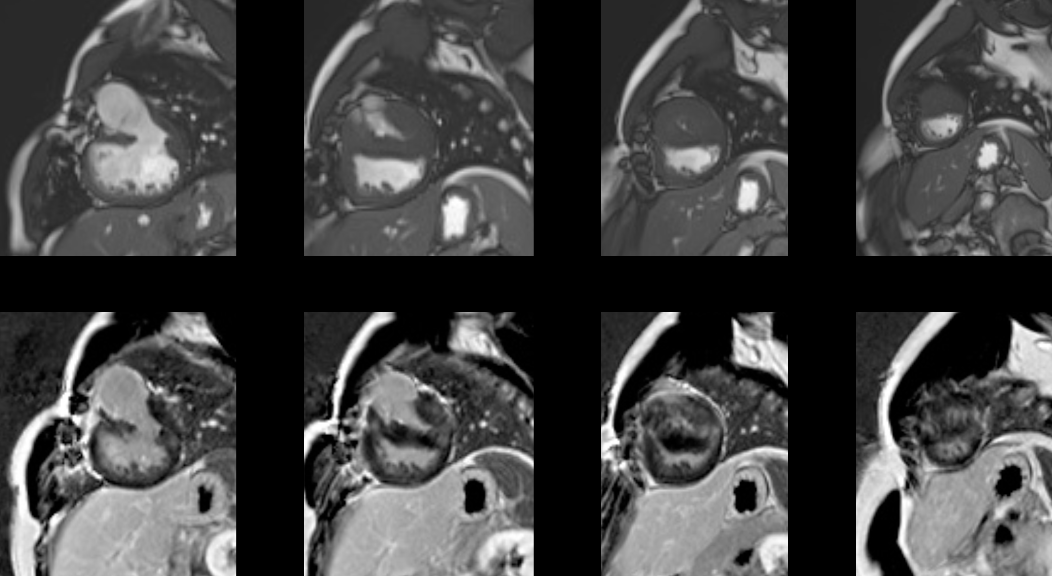
 Top row: four short-axis cine-SSFP slices. The first slice shows a wide interventricular communication. In the first and second slices, the aorta arises from the vestigial right ventricle. Hypertrophy of the interventricular septum and the free wall of the vestigial RV is evident in all slices. Bottom row: late gadolinium enhancement images. In the second and third slices, intramyocardial enhancement is visible at the RV insertion points in the interventricular septum. In the third and fourth slices, diffuse fibrosis of the vestigial RV free wall is observed.