Quick Fire Session
.jpg "Ronak Naik, MD photo")
Ronak Naik, MD
Associate Professor
Le Bonheur Children's Hospital, University of Tennessee Health Science Center
Memphis, Tennessee, United States
Ronak Naik, MD
Associate Professor
Le Bonheur Children's Hospital, University of Tennessee Health Science Center
Memphis, Tennessee, United States
Andrew Corson, MD
Pediatric Cardiology Fellow
Le Bonheur Children's Hospital
Germantown, Tennessee, United States

Anthony Merlocco, MD, MA, FSCMR
Director of Cardiac MRI
LeBonheur Children's Hospital
Memphis, Tennessee, United States
Nirbhay Parashar, MD
Pediatric Cardiology
Le Bonheur Children's Hospital
Jackson, Tennessee, United States
Sarah Parkerson, MD
Pediatric Cardiology Fellow, University of Tennessee Health Sciences Center
Le Bonheur Children's Hospital, United States

Jason N. Johnson, MD
Chief, Pediatric Cardiology
Le Bonheur Children's Hospital
Memphis, Tennessee, United States
 Figure 2: A. T2Weighted image short axis of LV showing increased hyperintensity (arrow). B. Post contrast Modified Look-Locker sequence showing area devoid of contrast uptake (arrow).
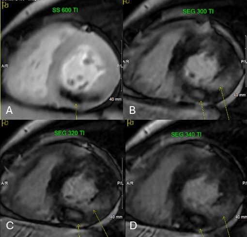
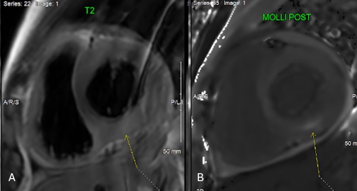 Figure 3: A. Inversion recovery image at higher inversion time of 600 msec showing area of microvascular obstruction (arrow) within the myocardial wall. Same area at usual inversion time of 300 msec (B), 320 msec (C) and 340 msec (D) (small arrow) showing well demarcated region of hyperintensity suggesting no contrast uptake. The long arrows indicate subendocardial to near transmural late gadolinium enhancement.