SCMR - ISMRM Co-Provided Workshop
CMR In Patients With Cardiac Implantable Electronic Devices: Optimizing Workflows And Image Quality.
The workshop is included in the registration rates.
|
|
 |
 |
|
Daniel Kim Co-Chair |
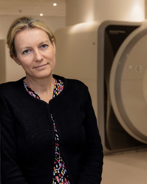
Charlotte Manisty Co-Chair |
Organizing Committee:
Kenneth Bilchick (United States), Harold Litt (United States), Aurelien Bustin (France), Peng Hu (United States), Anna Giulia Pavon (Switzerland), Sonya Narayan (United Kingdom)
Patients with cardiac implantable electronic devices (CIEDs) have a clear clinical need for high quality cardiac imaging, including tissue characterization, to both understand the aetiology and mechanisms of arrhythmias, and guide management strategies including electrophysiological interventions. Whilst historically MRI in patients with CIEDs was contraindicated, efforts from manufacturers has led to development of MR Conditional versions of most CIED systems, and studies in legacy MR Unlabelled devices have evidenced their relative safety with MRI provided appropriate protocols are followed. One residual challenge is imaging artifact arising from the CIED generator that limits image quality – particularly for tissue characterization sequences, leading many imagers to avoid CMR in patients with implantable cardioverter defibrillators (ICDs). This has resulted in inequity of provision and access to CMR for many patients with CIEDs.
Several academic groups have developed metal artifact mitigation strategies for improving image quality, and scanner manufacturers have adapted sequences to reduce the impact of artifact. However there are still concerns related to both the safety of CMR in patients with certain devices, and how to enable diagnostic imaging across all CMR sequences for patients with CIEDs.
This workshop will review and discuss major safety data, protocols, sequence adaptation and recent developments in CMR for patients with CIEDs.
Offered on Wednesday, February 4, the workshop seeks to provide an in-depth review of topics related to:
- Clinical indications for CMR in patients with CIEDs
- Safety of cardiac implantable electronic devices (CIEDs) – critical appraisal of the evidence and considerations with MR Unlabelled devices.
- Workflows for efficient CIED scanning (including monitoring)
- Fundamentals of imaging artifact – including both arrhythmia and device related considerations
- Artifact mitigation strategies – practical tips, novel sequences and WIPs
- Post processing optimisation including the application of AI
- Considerations for stress perfusion imaging in patients with CIEDs
- CMR guided VT ablation. Legal, regulatory and reimbursement challenges for CMR in CIED patients
- Future advancements: how can we bring imaging scientists and clinicians together with CIED and scanner manufacturers to advance the field?


